. 2008 Jul;15(1):62-73.
doi: 10.1016/j.devcel.2008.05.014.

Two distinct modes of ESCRT-III recognition are required for VPS4 functions in lysosomal protein targeting and HIV-1 budding

Affiliations

Two distinct modes of ESCRT-III recognition are required for VPS4 functions in lysosomal protein targeting and HIV-1 budding

Collin Kieffer et al. Dev Cell. 2008 Jul.

Abstract

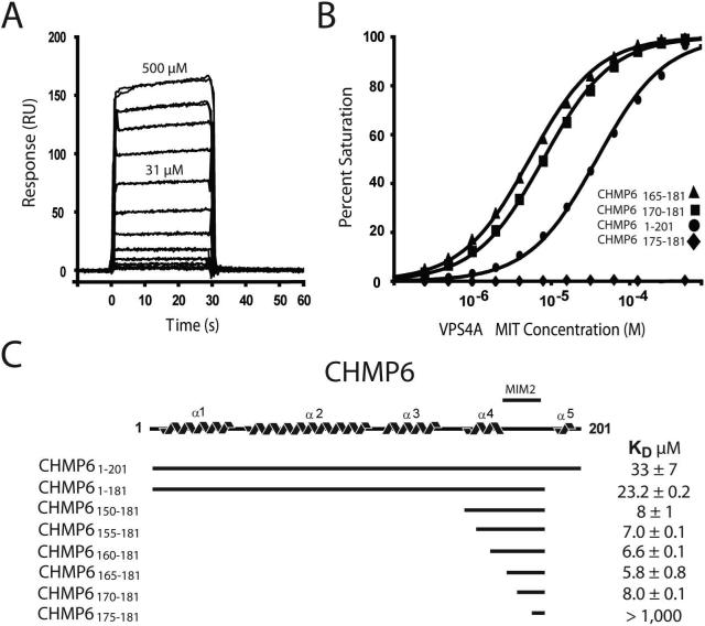
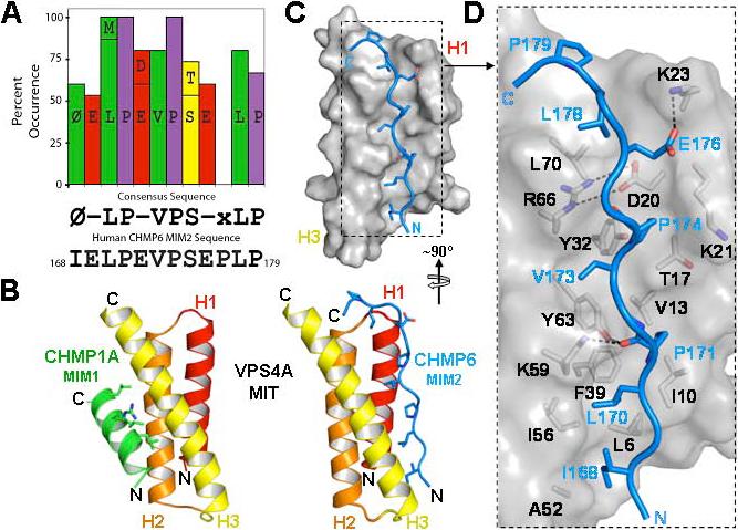
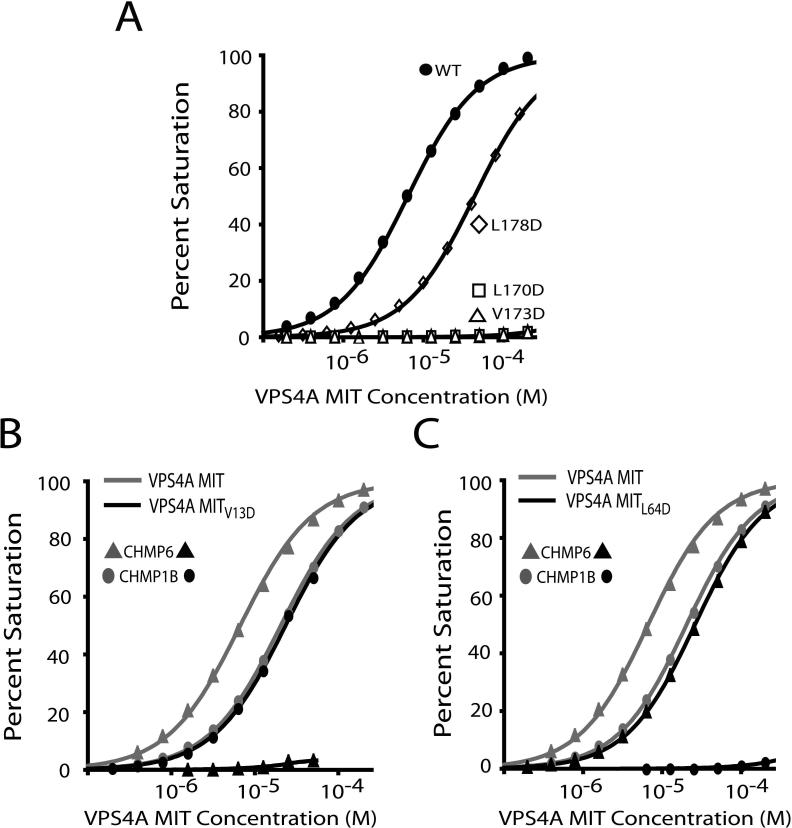
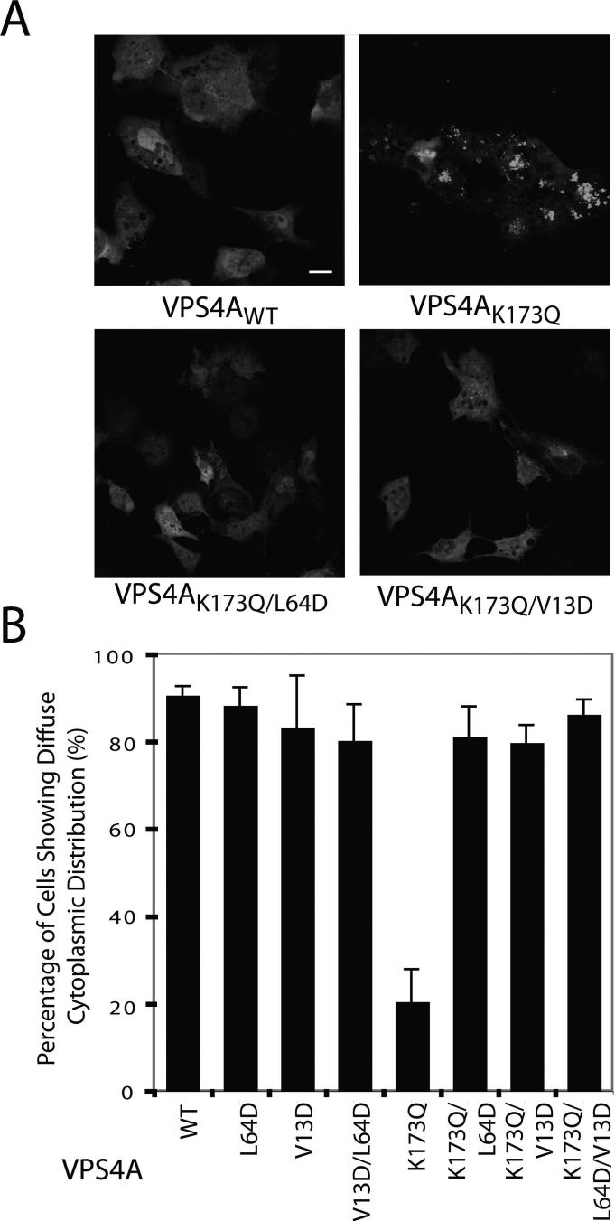
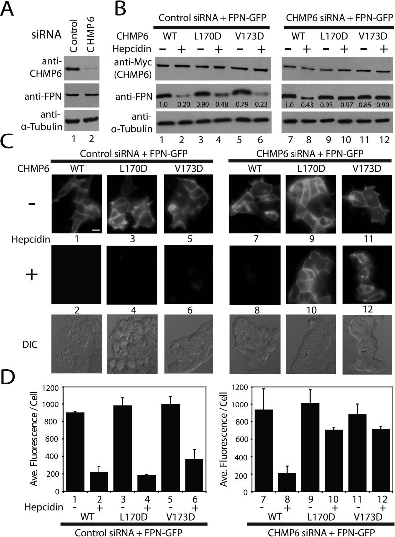
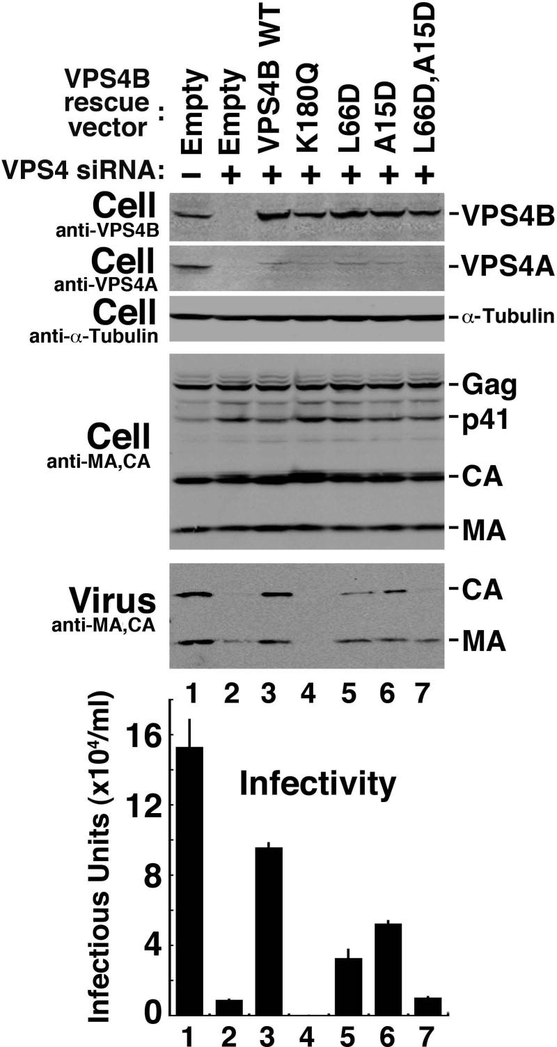
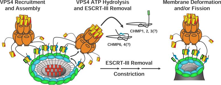
The ESCRT pathway mediates membrane remodeling during enveloped virus budding, cytokinesis, and intralumenal endosomal vesicle formation. Late in the pathway, a subset of membrane-associated ESCRT-III proteins display terminal amphipathic "MIM1" helices that bind and recruit VPS4 ATPases via their MIT domains. We now report that VPS4 MIT domains also bind a second, "MIM2" motif found in a different subset of ESCRT-III subunits. The solution structure of the VPS4 MIT-CHMP6 MIM2 complex revealed that MIM2 elements bind in extended conformations along the groove between the first and third helices of the MIT domain. Mutations that block VPS4 MIT-MIM2 interactions inhibit VPS4 recruitment, lysosomal protein targeting, and HIV-1 budding. MIT-MIM2 interactions appear to be common throughout the ESCRT pathway and possibly elsewhere, and we suggest how these interactions could contribute to a mechanism in which VPS4 and ESCRT-III proteins function together to constrict the necks of budding vesicles.


Figures

Figure 1
Figure 1. VPS4A MIT Binds a Unique Internal CHM6 MIT Interacting Motif (MIM2)
(A) Biosensorgrams showing concentration-dependent binding of purified VPS4A MIT (residues 1−84, 0−500 μM) to full length GST-CHMP61−201. (B) Biosensor binding isotherms showing VPS4A MIT binding to a series of different GST-CHMP6 deletion mutants (see inset key). Binding to a GST control surface was negligible (not shown). (C) Schematic illustration of the predicted CHMP6 structure, indicating the location of the CHMP6 MIM2 between helices α4 and α5. Dissociation constants for the different CHMP6 constructs are shown at right.
Figure 2
Figure 2. Solution Structure of the VPS4A MIT-CHMP6 MIM2 Complex
(A) Graphic summary showing MIM2 residues that are >50% conserved across eukaryotic CHMP6 proteins (and see Supplemental Table 2), the resulting MIM2 consensus sequence, and the CHMP6 MIM2 sequence. “Φ” denotes an aliphatic residue and “-“ denotes acidic residues. (B) Structures of human VPS4A MIT in complex with the MIM1 element from CHMP1A (left structure, MIM1 helix in green) and the CHMP6 MIM2 element (right, MIM2 strand in blue). The MIT domains are shown in the same orientation to emphasize that the MIM1 and MIM2 elements bind in different grooves of the MIT three helix bundle. Key MIM contact residues are shown explicitly in both structures. (C, D) Overview (C) and detailed (D) views of the VPS4A MIT domain (gray and black, space filling representation) in complex with the CHMP6 MIM2 element (stick representation, blue). Key side chains on both sides of the interface are shown in (D), with selected intermolecular salt bridges and hydrogen bonds indicated by dashed lines.
Figure 3
Figure 3. Mutational Analyses of the VPS4A MIT-CHMP6 Complex
(A) Biosensor binding isotherms showing VPS4A MIT binding to wild type and mutant CHMP6 MIM2 peptides (labeled). Note that the CHMP6 L178D mutation reduces the binding affinity ∼8-fold (from KD=5.8±0.8 to KD=44.4±0.2 μM), whereas the L170D and Vl173D mutations block all detectable binding. (B) Biosensor binding isotherms showing that the V13D mutation in VPS4A MIT helix 1 blocks CHMP6 MIM2 binding (triangles) but does not affect CHMP1B MIM1 binding (ovals). (C) Biosensor binding isotherms showing that the L64D mutation in VPS4A MIT helix 3 blocks CHMP1B MIM1 binding (ovals), but has much less effect on CHMP6 MIM2 binding (triangles), altering CHMP6 MIM2 binding from KD = 5.8 ± 0.8 μM to KD = 26.1 ± 0.8 μM.
Figure 4
Figure 4. VPS4A MIT Mutations that Block MIM1 and MIM2 Binding also Inhibit Recruitment of VPS4AK173Q to Class E Endosomal Compartments
A) Confocal fluorescence slices showing cellular distributions of wild type GFP–VPS4A (VPSAWT, upper left, negative control) and GFP-VPS4A proteins with the following mutations: K173Q (upper right, positive control, mutation blocks ATP binding and induces class E compartment formation), K173Q,L64D (lower left, K173Q mutation blocks ATP binding and L64D mutation blocks MIM1 binding), and K173Q,V13D (lower right, K173Q mutation blocks ATP binding and V13D mutation blocks MIM2 binding). Scale bar is 20 μm. (B) Quantification of cells showing diffuse cytoplasmic localization of wild type and mutant GFP-VPS4A proteins (mutations are listed below). Two blinded sets of >100 cells of each type were scored, and error bars indicate standard deviations. Note that the VPS4A MIT L64D and V13D both mutations alleviate the Class E compartment recruitment induced by the K173Q mutation (compare lanes 6−8 to lane 5).
Figure 5
Figure 5. CHMP6 MIM2 Mutations Inhibit Hepcidin-induced Lysosomal Degradation of Ferroportin
(A) Western blot analyses showing efficient siRNA depletion of endogenous CHMP6 protein (top blot, compare lanes 1 and 2), without changes in ferroportin–GFP (FPN) or α–Tubulin levels (bottom two blots). (B) Western blot analyses of hepcidin–induced ferroportin–GFP downregulation in the presence of wild type and mutant CHMP6 proteins. Left panels (controls) show ferroportin levels (middle blot) in the absence (−) or presence (+) of hepcidin treatment of cells expressing wild type and mutant CHMP6-Myc proteins (top blot) in the presence of endogenous CHMP6 (control siRNA). Right panels show the same experiment, but with depletion of endogenous CHMP6, so that only the exogenous, siRNA-resistant CHMP6-Myc proteins were present. Integrated intensities were normalized to the untreated control and are shown below each band. Note that ferroportin–GFP levels decreased 2.3–fold upon hepcidin treatment in the presence of wild type CHMP6-Myc (compare lanes 7 and 8) but did not decrease in the presence of CHMP6-Myc proteins with inactivating MIM2 point mutations (L170D and V173D). (C, D) Epifluorescence and differential interference contrast (DIC) images (C) and quantification (D) showing the effects of hepcidin treatment and CHMP6 mutations on ferroportin-GFP levels. Samples 1−12 correspond to the equivalent samples in part (B). Note that the data again show that wild type CHMP6-Myc supports efficient hepcidin-induced ferroportin-GFP downregulation (compare samples 7 and 8, 4.2-fold reduction), whereas the Myc-CHMP6 MIM2 mutants do not (compare samples 9 and 11 to 10 and 12). Scale bar is 20 μm, and DIC images of the hepcidin-treated cells are provided for reference in the lower panels of part (C). Quantifications presented in (D) show the average fluorescence/cell from two data sets (one blinded) of >100 cells each, and error bars indicate standard deviations.
Figure 6
Figure 6. The VPS4B MIT MIM1 and MIM2 Binding Surfaces Both Contribute to Efficient HIV-1 Budding and Replication
Western blots 1 and 2 show simultaneous siRNA depletion of endogenous VPS4A and VPS4B (Compare lanes 1 and 2) and re-expression of exogenous siRNA-resistant wild type and mutant VPS4B proteins (blot 1, Lanes 3−7). Western blot 3 shows an α–Tubulin loading control. Western blots 4 and 5 show levels of the viral Gag protein and its processed MA and CA products in the cytoplasm (Cell, panel 4, note that all cells were transfected with a proviral HIV-1 expression construct), and released as viral particles (Virus). Panel 6 shows viral titers, as assayed in single cycle replication assays of the culture supernatants. The experiment shows that simultaneous depletion of both human VPS4 isoforms blocks HIV-1 release and infectivity (panels 5 and 6, compare lanes 1 and 2), that reintroduction of a wild type, siRNA-resistant VPS4B protein construct significantly rescues virus release and titer (compare lanes 2 and 3), and that virus release and titer are inhibited by the VPS4B K180Q (lane 4, positive control), L66D (lane 5, inhibited MIM1 binding), A15D (lane 6, inhibited MIM2 binding) and L66D,A15D (lane 7, inhibited MIM1 and MIM2 binding).
Figure 7
Figure 7. Model for VPS4 Recruitment and ESCRT-III Filament Constriction
Schematic model showing how different ESCRT-III subunits (blue and green) could co-assemble into concentric rings that display C-terminal MIM1 (green) and internal MIM2 elements (blue), thereby creating a high affinity VPS4 binding surface (orange, with the three helices of the MIT domains in red, orange, and yellow, respectively). Recruited VPS4 ATPases could then remove individual ESCRT-III subunits, constricting the rings about cargoes (red) and thereby helping drive vesicle extrusion and neck closure (see text for full details). We note that six molecules of the Vps4 activator, LIP5/Vta1p, also bind the VPS4 beta domains and make additional MIT-ESCRT-III interactions, but for clarity these additional interactions are not shown.

Comment in

  • Findings of Research Misconduct.
    [No authors listed] [No authors listed] Fed Regist. 2023 Sep 5;88(170):60694-60695. Fed Regist. 2023. PMID: 37736265 Free PMC article. No abstract available.

References

    1. Azmi I, Davies B, Dimaano C, Payne J, Eckert D, Babst M, Katzmann DJ. Recycling of ESCRTs by the AAA-ATPase Vps4 is regulated by a conserved VSL region in Vta1. J Cell Biol. 2006;172:705–717. - PMC - PubMed
    1. Azmi IF, Davies BA, Xiao J, Babst M, Xu Z, Katzmann DJ. ESCRT-III Family Members Stimulate Vps4 ATPase Activity Directly or via Vta1. Dev Cell. 2008;14:50–61. - PubMed
    1. Babst M, Katzmann D, Estepa-Sabal E, Meerloo T, Emr S. Escrt-III. An endosome-associated heterooligomeric protein complex required for mvb sorting. Dev Cell. 2002;3:271–282. - PubMed
    1. Babst M, Wendland B, Estepa EJ, Emr SD. The Vps4p AAA ATPase regulates membrane association of a Vps protein complex required for normal endosome function. EMBO J. 1998;17:2982–2993. - PMC - PubMed
    1. Bajorek M, Morita E, Skalicky JJ, Morham SG, Babst M, Sundquist WI. Biochemical Analyses of Human IST1 and Its Function in Cytokinesis. 2008 submitted. - PMC - PubMed

Publication types

MeSH terms

Substances