. 2008 Jul;15(1):134-45.
doi: 10.1016/j.devcel.2008.05.001.

The chirality of gut rotation derives from left-right asymmetric changes in the architecture of the dorsal mesentery

Affiliations

The chirality of gut rotation derives from left-right asymmetric changes in the architecture of the dorsal mesentery

Nicole M Davis et al. Dev Cell. 2008 Jul.

Abstract

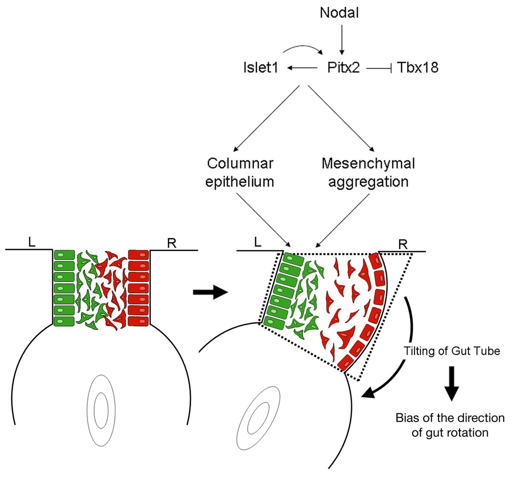
We have investigated the structural basis by which the counterclockwise direction of the amniote gut is established. The chirality of midgut looping is determined by left-right asymmetries in the cellular architecture of the dorsal mesentery, the structure that connects the primitive gut tube to the body wall. The mesenchymal cells of the dorsal mesentery are more condensed on the left side than on the right and, additionally, the overlying epithelium on the left side exhibits a columnar morphology, in contrast to a cuboidal morphology on the right. These properties are instructed by a set of transcription factors: Pitx2 and Isl1 specifically expressed on the left side, and Tbx18 expressed on the right, regulated downstream of the secreted protein Nodal which is present exclusively on the left side. The resultant differences in cellular organization cause the mesentery to assume a trapezoidal shape, tilting the primitive gut tube leftward.


Figures

Figure 1
Figure 1. The formation and rotation of the gut tube
(A) Cross-section during gut tube formation. (B) Side view of the primitive gut tube. (C) Cross-section of the primitive gut tube. (D) Primary intestinal loop moving into the umbilicus (in mammals) or yolk sac (in birds). (E) Gut tube retracting into the body cavity. (F) Final orientation of the midgut‥ Endoderm (yellow), mesoderm (pink).
Figure 2
Figure 2. Morphological asymmetry in the midgut dorsal mesentery regulated by Pitx2 and Isl1
(A–C) Histology of cross-section of wild type midgut midgut‥ (D–F) Cross-section of midgut following misexpression of Isl1. (G–I) Cross-section of midgut following misexpression of Pitx2. (J) Quantification of the extent of midgut tilting in uninfected and infected embryos. Each line represents a single embryo, either wild-type or infected with Pix2 or Isl1 virus, as indicated. Representative sections were measured every 25 microns. All sections shown with the embryo’s left side oriented on the left (applies to all figures).
Figure 3
Figure 3. Cellular asymmetries in the dorsal mesentery
Columnar (A) versus cuboidal (B) cell morphology of the epithelium in the dorsal mesentery as revealed by transverse section and H&E staining at HH stage 20. (C) Phalloidin staining (red) of transverse section of the dorsal mesentery; DAPI (blue) counterstain. (D–E) In ovo gfp-electroporation of left and right side of the dorsal mesentery at HH stage 14. Electroporated dorsal mesentery (green) harvested at 12h (D) or 36h (E) after manipulation. Phalloidin (red) counterstain. (F) Confocal microscopy of electroporated right dorsal mesentery harvested 24 hours after manipulation; posterior is left, anterior is right, dorsal is up and ventral is down. High magnification (63X) of columnar epithelial cells on the left (G–I) and on the right (M–O) side of the dorsal mesentery (green, GFP; red, phalloidin, blue, β-catenin). Transmission electron microscopy of left (J–L) and right (P–R) side of the dorsal mesentery. Underlying basement membrane false-colored purple. (P,Q) Epithelial cells on the right. Higher magnification electron microscopy (23,000X) reveals tight junctions and desmosomes in the left (L) and right (R) epithelia of the dorsal mesentery.
Figure 4
Figure 4. Analysis of changes in epithelial cell shape and condensation of the mesenchyme
(A–F) Quantitative analyses of the epithelial cell height and width on the left and right sides of the wild type dorsal mesentery and following misexpression of either Pitx2 or Isl1. Error bars show SD. (A) Height of left versus right epithelial cells at mid-caudal level. (B) Width of left versus right epithelial cells at mid-caudal level. (C) Height of left versus right epithelial cells following misexpression of either Pitx2 or Isl1 at mid-caudal 32 level. (D) Width of left versus right epithelial cells following misexpression of either Pitx2 or Isl1 at mid-caudal level. (E) Height of left versus right epithelial cells at midrostral level. (F) Width of left versus right epithelial cells at mid-rostral level. (G) Dorsal mesentery prior to tilting. (H) Dorsal mesentery at Stage 22. (I) Cell counts of left and right DAPI-positive mesenchymal cells (blue). (J) Immunohistochemistry to phospho-Histone H3 (red). (K) TUNEL assay (red) reveals no apoptotic cells in the midgut (inset mesonephric duct). (L) DAPI.
Figure 5
Figure 5. Pitx2 is necessary as well as sufficient for specifying the unique morphology and gene expression pattern on the left side of the dorsal mesentery
(A) Wild type mouse dorsal mesentery at E10.75. Red lines, height of epithelia. (B) Dorsal mesentery in E10.75 mouse embryos deficient for Pitx2. (C) Wild type Isl1 expression at E10.75 in dorsal mesentery and in neural tube (inset). (D) Isl1 expression in Pitx2-deficient embryos. (E) Tbx18 expression in wild type E10.75 mouse. (F) Expression of Tbx18 in the absence of Pitx2.
Figure 6
Figure 6. Asymmetric expression of transcription factors in the dorsal mesentery is regulated by the Nodal-Pitx2 pathway
(A–C) Section in situ hybridization at HH stage 21 reveals asymmetric expression of Pitx2 (A), Isl1 (B), and Tbx18 (C) in the dorsal mesentery at the level of the midgut. (D) Section stained with antibodies (3C2) recognizing a retroviral coat protein. Expression of Pitx2 (E) Isl1 (F) and Tbx18 (G) following Nodal misexpression at HH stage 4. Expression of Isl1 (H) and Tbx18 (I) following Pitx2 misexpression.. Expression of Pitx2 (J) and Tbx18 (K) following Isl1 misexpression. (L) Whole mount of wild type gut at HH stage 22. (M) Gut following Isl1 misexpression. (N) Histology of wild type gut .(O) Histology following misexpression of Pitx2.
Figure 7
Figure 7
Model for the directional looping of the gut tube.

References

    1. Bamforth SD, Braganca J, Farthing CR, Schneider JE, Broadbent C, Michell AC, Clarke K, Neubauer S, Norris D, Brown NA, et al. Cited2 controls left-right patterning and heart development through a Nodal-Pitx2c pathway. Nat Genet. 2004;36:1189–1196. - PubMed
    1. Begemann G, Gibert Y, Meyer A, Ingham PW. Cloning of zebrafish T-box genes tbx15 and tbx18 and their expression during embryonic development. Mech Dev. 2002;114:137–141. - PubMed
    1. Brent AE, Schweitzer R, Tabin CJ. A somitic compartment of tendon progenitors. Cell. 113:235–248. - PubMed
    1. Campione M, Steinbeisser H, Schweickert A, Deissler K, van Bebber F, Lowe LA, Nowotschin S, Viebahn C, Haffter P, Kuehn MR, et al. The homeobox gene Pitx2: mediator of asymmetric left-right signaling in vertebrate heart and gut looping. Development. 1999;126:1225–1234. - PubMed
    1. Collignon J, Varlet I, Robertson EJ. Relationship between asymmetric nodal expression and the direction of embryonic turning. Nature. 1996;381:155–158. - PubMed

Publication types

MeSH terms