. 2008 Nov;29(6):971-7.
doi: 10.1016/j.neuro.2008.06.003. Epub 2008 Jun 19.

Different effects of selective dopamine uptake inhibitors, GBR 12909 and WIN 35428, on HIV-1 Tat toxicity in rat fetal midbrain neurons

Affiliations

Different effects of selective dopamine uptake inhibitors, GBR 12909 and WIN 35428, on HIV-1 Tat toxicity in rat fetal midbrain neurons

Michael Y Aksenov et al. Neurotoxicology. 2008 Nov.

Abstract

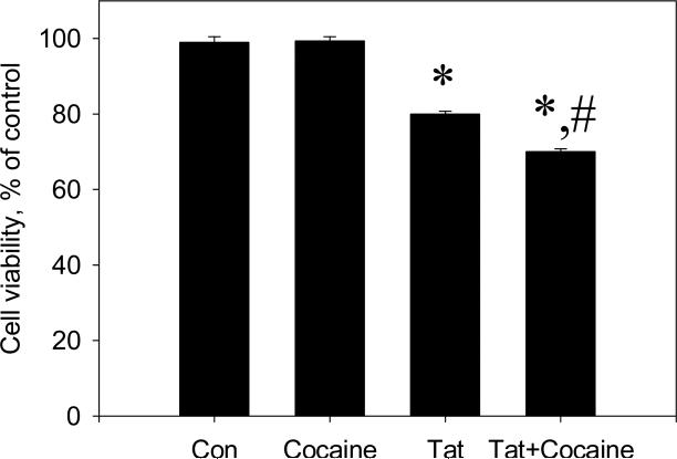
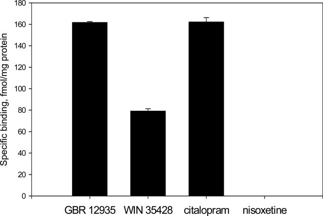
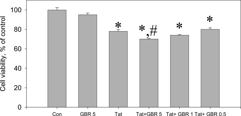
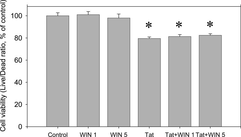
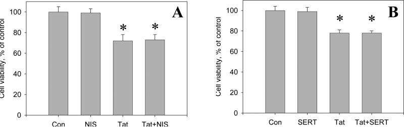
Drug abuse is a risk factor for neurological complications in HIV infection. Cocaine has been shown to exacerbate HIV-associated brain pathology and enhance neurotoxicity of HIV-1 Tat and gp120 proteins. In this study, we found that the selective inhibitor of dopamine transporter (DAT) function, 1-[2-[bis(4-fluorophenyl) methoxy]ethyl]-4-(3-phenylpropyl) piperazine (GBR 12909, vanoxerine), but not the selective inhibitors of serotonin and norepinephrine (SERT and NET) transporters, sertraline and nizoxetine, emulated cocaine-mediated enhancement of Tat neurotoxicity in rat fetal midbrain primary cell cultures. Similar to cocaine, the significant increase of Tat toxicity in midbrain cell cultures was observed at micromolar dose (5microM) of GBR 12909. However, different doses of another selective dopamine uptake inhibitor, WIN 35428 did not affect Tat neurotoxicity. The study supports the hypothesis that changes in control of dopamine (DA) homeostasis are important for the cocaine-mediated enhancement of HIV-1 Tat neurotoxicity. Our results also demonstrate that inhibitors of DA uptake, which can bind to different domains of DAT, differ in their ability to mimic synergistic toxicity of cocaine and HIV-1 Tat in the midbrain cell culture.


Figures

Figure 1
Figure 1
The effect of cocaine on the toxicity of Tat 1-86 in primary rat fetal midbrain cell culture. Graph represents relative (compared to non-treated controls) changes in Live/Dead ratios 48 hours following the addition of 50 nM Tat 1-86 or 50 nM Tat 1-86 + 1.5 μM cocaine to the cell cultures. Data presented as mean values ± SEM, n of sister cultures analyzed = 7-14 per each variant of treatment. *- marks significant (P<0.05) difference between Tat-treated (50 nM Tat) group and non-treated control group; *,#-marks that this experimental group (50 nM Tat+ 1.5 μM cocaine) was significantly (P<0.05) different from non-treated control (*) and from Tat-treated group (#).
Figure 2
Figure 2
The specific binding of DAT, SERT, and NET-selective ligands in rat fetal midbrain cell cultures. Data presented as mean values ± SEM, n of sister cultures analyzed = 4 per each ligand.
Figure 3
Figure 3
Representative images of anti-DAT and anti-SERT immunoreactivity in rat fetal midbrain cell cultures. (A) The image of anti-DAT/Hoechst staining in rat fetal midbrain cell cultures shows positive red immunofluorescence in neuronal somata and processes in 12th DIV midbrain neurons. Hoechst blue fluorescence counter stains cell nuclei. Red background fluorescence was insignificant in “no-primary antibody” controls (image not shown). (B) The image of anti-SERT/Hoechst staining in rat midbrain cell cultures shows positive anti-SERT (green) immunofluorescence in 12th DIV midbrain neurons. Hoechst staining is used to counter stain cell nuclei. Green background fluorescence was insignificant in “no-primary antibody” controls (image not shown).
Figure 3
Figure 3
Representative images of anti-DAT and anti-SERT immunoreactivity in rat fetal midbrain cell cultures. (A) The image of anti-DAT/Hoechst staining in rat fetal midbrain cell cultures shows positive red immunofluorescence in neuronal somata and processes in 12th DIV midbrain neurons. Hoechst blue fluorescence counter stains cell nuclei. Red background fluorescence was insignificant in “no-primary antibody” controls (image not shown). (B) The image of anti-SERT/Hoechst staining in rat midbrain cell cultures shows positive anti-SERT (green) immunofluorescence in 12th DIV midbrain neurons. Hoechst staining is used to counter stain cell nuclei. Green background fluorescence was insignificant in “no-primary antibody” controls (image not shown).
Figure 4
Figure 4
The effect of the selective dopamine transporter inhibitor, GBR 12909, on the toxicity of Tat 1-86 in primary rat fetal midbrain cell culture. Results presented as mean % of Calcein/Ethidium bromide fluorescence (Live/Dead ratio)vs non-treated control ± SEM, n of sister cultures analyzed = 7-13 per each variant of treatment. *- marks significant (P<0.05) difference between the treated group (50 nM Tat or 50 nM Tat + non-toxic dose of GBR 12909) and non-treated control group; *,#-marks that this experimental group (50 nM Tat+ non-toxic dose of GBR 12909) was significantly (P<0.05) different from non-treated control (*) and from Tat-treated group (#). Similar to cocaine, 5 μM GBR 12909 significantly enhances neurotoxicity of 50 nM Tat following 48 hour-exposure. All doses of GBR 12909 used in these experiments (0.5, 1, or 5 μM) did not cause changes of Live/Dead ratios in rat fetal midbrain cell cultures. Cell viability of a group of cultures exposed to 5 μM GBR 12909 is included in the graph.
Figure 5
Figure 5
The effect of the selective DAT inhibitor, WIN 35428, on the toxicity of Tat 1-86 in primary rat fetal midbrain cell cultures. Results presented as mean % of Calcein/Ethidium bromide fluorescence (Live/Dead ratio)vs non-treated control ± SEM, n of sister cultures analyzed = 7-17 per each variant of treatment. Two different doses of WIN 35428 (1 and 5 μM) used in these experiments were not toxic to cultured rat fetal midbrain neurons and did not enhance the toxicity of Tat 1-86 determined after 48 hour-exposure. *- marks the significant (P<0.05) difference between Tat-treated group or Tat+WIN 35428 –treated group and non-treated control group;
Figure 6
Figure 6
The effect of the selective norepinephrine transporter inhibitor, nisoxetine (A), and the selective serotonine transporter inhibitor, sertraline (B) on the toxicity of Tat 1-86 in primary rat fetal midbrain cell culture. Results presented as mean % of Calcein/Ethidium bromide fluorescence (Live/Dead ratio)vs non-treated control ± SEM, n of sister cultures analyzed = 6-12 per each variant of treatment. *- marks significant (P<0.05) difference between the treated group (50 nM Tat or 50 nM Tat + non-toxic dose of sertraline / nisoxetine) and non-treated control group.

References

    1. Aksenov MY, Hasselrot U, Wu G, Nath A, Anderson C, Mactutus CF, Booze RM. Temporal relationships between HIV-1 Tat-induced neuronal degeneration, OX-42 immunoreactivity, reactive astrocytosis, and protein oxidation in the rat striatum. Brain Res. 2003;987:1–9. - PubMed
    1. Aksenov MY, Aksenova MV, Nath A, Ray PD, Mactutus CF, Booze RM. Cocaine-mediated enhancement of Tat toxicity in rat hippocampal cell cultures: the role of oxidative stress and D1 dopamine receptor. Neurotox. 2006;27:217–228. - PubMed
    1. Aksenova MV, Silvers JM, Aksenov MY, Nath A, Ray PD, Mactutus CF, Booze RM. HIV-1 Tat neurotoxicity in primary cultures of rat midbrain fetal neurons: changes in dopamine transporter binding and immunoreactivity. Neurosci Lett. 2006;395:235–239. - PubMed
    1. Blakely RD, De Felice LJ, Hartzell HC. Molecular physiology of norepinephrine and serotonin transporters. J. Exp. Biol. 1994;196:263–281. - PubMed
    1. Boos TL, Greiner E, Calhoun WJ, Prisinzano TE, Nightingale B, Dersch CM, Rothman RB, Jacobson AE, Rice KC. Structure–activity relationships of substituted N-benzyl piperidines in the GBR series: Synthesis of 4-(2-(bis(4-fluorophenyl) methoxy)ethyl)-1-(2-trifluoromethylbenzyl) piperidine, an allosteric modulator of the serotonin transporter. Bioorganic & Medicinal Chemistry. 2006;14:3967–3973. - PubMed

MeSH terms

Substances

LinkOut - more resources