Review
. 2008 Aug;24(8):398-407.
doi: 10.1016/j.tig.2008.05.008. Epub 2008 Jul 5.

Iron-sulfur cluster biogenesis and human disease

Affiliations
Review

Iron-sulfur cluster biogenesis and human disease

Tracey A Rouault et al. Trends Genet. 2008 Aug.

Abstract

Iron-sulfur (Fe-S) clusters are essential for numerous biological processes, including mitochondrial respiratory chain activity and various other enzymatic and regulatory functions. Human Fe-S cluster assembly proteins are frequently encoded by single genes, and inherited defects in some of these genes cause disease. Recently, the spectrum of diseases attributable to abnormal Fe-S cluster biogenesis has extended beyond Friedreich ataxia to include a sideroblastic anemia with deficiency of glutaredoxin 5 and a myopathy associated with a deficiency of a Fe-S cluster assembly scaffold protein, ISCU. Mutations within other mammalian Fe-S cluster assembly genes could be causative for human diseases that manifest distinctive combinations of tissue-specific impairments. Thus, defects in the iron-sulfur cluster biogenesis pathway could underlie many human diseases.


Figures

Figure I
Figure I
Mitochondrial iron overload in Friedreich ataxia, a myopathy with ISCU deficiency, and an acquired siderblastic anemia. Mitochondrial overload was detected in patient samples using histochemical stain and electron microscopy. Blue arrows indicate mitochondrial iron deposits and purple arrows indicate well preserved cristae and a relative absence of iron deposits. Prussian blue staining revealed that excess ferric iron was present in the hearts of a 9-year-old patient (b) and a 26-year-old patient (c) with FRDA compared with the heart of a unaffected 35-year-old individual (a). Biochemical and electron microscopic analyses have previously indicated that iron accumulates mainly in the mitochondria. In normal skeletal muscle biopsies, absence of brown DAB-enhanced Prussian blue staining is indicative of normal levels of ferric iron (d), whereas brown punctate deposits reveal mitochondrial iron overload throughout the individual muscle fibers of patients with myopathy and ISCU deficiency (e), a finding that has been confirmed by electron microscopy (f) [54]. In human sideroblastic anemias, dark deposits within distorted mitochondria are caused by iron overload [64]. Electron microscopy analysis of normoblasts (normal red cell precursors) of a patient from an early stage in development of acquired sideroblastic anemia showed that most of the mitochondria were still well preserved (as evidenced by well preserved cristae) (g), whereas in the sideroblasts at later stages, the mitochondria became distorted and increasingly loaded with iron, which appears as black deposits within the matrix (h and i).
Figure 1
Figure 1
Disruption of iron–sulfur (Fe–S) proteins can have a myriad of deleterious cellular consequences. Because Fe–S clusters are essential electron carriers and enzyme cofactors in many proteins, defects in Fe–S clusters biogenesis can disrupt many cellular process. (a) Fe–S clusters are essential cofactors in energy metabolism. Fe–S proteins are among the most important electron carriers in nature and are particularly important in the mitochondrial respiratory chain, in which up to 12 different Fe–S clusters shuttle electrons through complexes I–III. Electrons from the oxidation of NADH and succinate are transferred through a chain of redox centers consist of flavins, Fe–S clusters, ubiquinone (Q and QH2), hemes and copper centers (CuA and CuB) to reduce O2 to H2O. The free energy of electron transport is coupled to ATP synthesis. In addition, the ability of Fe–S clusters to coordinate ligands and stabilize protein structures also allows them to facilitate various enzymatic functions. For instance, mitochondrial aconitase is an integral part of the tricarboxylic acid cycle (TCA cycle), and its [4 Fe–4S] cluster is essential for substrate binding and activation. (b) Because Fe–S proteins play a critical role in a wide range of cellular activities, mutations or pathological conditions that disrupt Fe–S cluster stability or biogenesis/repair are associated with several human diseases. For instance, germline mutations of the gene encoding succinate dehydrogenase subunit B (SDHB), a Fe–S protein in respiratory complex II, are a major cause of cancer of the kidney, adrenal gland and thyroid gland [63]. Mutations that destabilize the Fe–S clusters in DNA repair enzymes XPD and FancJ are associated with the phenotypes in patients with trichothiodystrophy and Fanconi anemia respectively [3]. Severe defects in Fe–S cluster biogenesis/repair can profoundly decrease the activities of respiratory complexes, the TCA cycle and the heme biosynthesis pathway [–9], resulting in decreased energy production and increased oxidative stress. In addition, disruption of Fe–S cluster biogenesis can lead to mitochondrial iron overload and cytosolic iron depletion. In the cytosol, defects in Fe–S cluster biogenesis might affect cytosolic aconitase (c-aconitase) activity and therefore citrate metabolism, which can disrupt the balance between glycolysis and fatty acid biosynthesis. Defects in cytosolic Fe–S cluster biogenesis/repair might also impair ribosome biogenesis, and purine catabolism pathways.
Figure 2
Figure 2
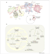
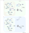
Iron–sulfur (Fe-S) cluster biogenesis in eukaryotes. Fe–S cluster biogenesis is a complex pathway involving many enzymes and proteins. (a) In the mitochondria and cytosol of mammalian cells, cysteine desulfurases (m-ISCS and c-ISCS) remove sulfur from free cysteine and donate the sulfur to scaffold proteins (e.g. m-ISCU and c-ISCU), whereas frataxin (FXN) is proposed to be an iron chaperone that delivers iron in an accessible form. Scaffold proteins can assemble and donate either [2Fe–2S] (not shown) or [4Fe–4S] clusters to recipient proteins. In the mitochondria, redox proteins such as FDX and FDXR likely deliver reducing equivalents needed for cluster assembly, and protein chaperones HSPA9/HSCB and glutaredoxin GRX5 facilitate the transfer of the cluster from scaffolds to recipient proteins. In the cytosol, additional factors including NUBP1, NUBP2, NARF and CIAO1 might facilitate cluster transfer. ISCS and ISCU are shown in ribbon structures. Other mitochondrial cluster biogenesis factors are shown in light blue ovals and cytoplasmic factors in dark blue ovals. (b) In yeast mitochondria, cysteine desulfurase Nfs1p, Isd11p, frataxin homolog Yfh1p and redox proteins Yah1p and Arh1p facilitate cluster assembly in scaffold proteins, whereas chaperone proteins Ssq1p and Jac1p and glutaredoxin Grx5p facilitate cluster transfer from scaffold to recipient proteins. The scaffold proteins Isu1p, Isu2p and Yfh1p have thus far only been detected in the mitochondria matrix, leading to the proposal that cluster assembly occurs exclusively in the mitochondria matrix, and preformed clusters are exported through Atm1p and transferred to cytosolic recipients through Cfd1p, Nar1p, Cia1p and Nbp35p. Nfs1p and Isu1p are shown in ribbon structures. Other mitochondrial cluster biogenesis factors are shown in light blue ovals and cytoplasmic factors in dark blue ovals.
Figure 3
Figure 3
Mutations in Friedreich ataxia, sideroblastic anemia with GLRX5 deficiency and myopathy with ISCU deficiency. In >98% of Friedreich ataxia (FRDA) cases, the defect is a GAA repeat expansion in the first intron of the Frataxin (FXN) gene. A small subset of patients have one expanded allele and a second allele harboring a premature stop codon or point mutation. In the patient with GLRX5 deficiency, a homologous silent mutation in exon 1 interferes with splicing and drastically reduces levels of fully processed mature GLRX5 mRNA. In myopathy with ISCU deficiency, a single G to C mutation in the fourth intron of ISCU activates a weak splice acceptor site and reduces the production of normal ISCU protein.

Similar articles

Cited by

References

    1. Johnson DC, et al. Structure, function, and formation of biological iron-sulfur clusters. Annu Rev Biochem. 2005;74:247–281. - PubMed
    1. Lill R, Muhlenhoff U. Maturation of iron-sulfur proteins in eukaryotes: mechanisms, connected processes, and diseases. Annu Rev Biochem. 2008;77:669–700. - PubMed
    1. Rudolf J, et al. The DNA repair helicases XPD and FancJ have essential iron-sulfur domains. Mol Cell. 2006;23:801–808. - PubMed
    1. Wallander ML, et al. Molecular control of vertebrate iron homeostasis by iron regulatory proteins. Biochim Biophys Acta. 2006;1763:668–689. - PMC - PubMed
    1. Rouault TA. The role of iron regulatory proteins in mammalian iron homeostasis and disease. Nat Chem Biol. 2006;2:406–414. - PubMed

Publication types