. 2008 Jul 15;181(2):1357-64.
doi: 10.4049/jimmunol.181.2.1357.

IL-17A inhibits the expansion of IL-17A-producing T cells in mice through "short-loop" inhibition via IL-17 receptor

Affiliations

IL-17A inhibits the expansion of IL-17A-producing T cells in mice through "short-loop" inhibition via IL-17 receptor

Emily Smith et al. J Immunol. .

Abstract

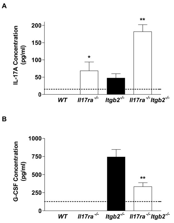
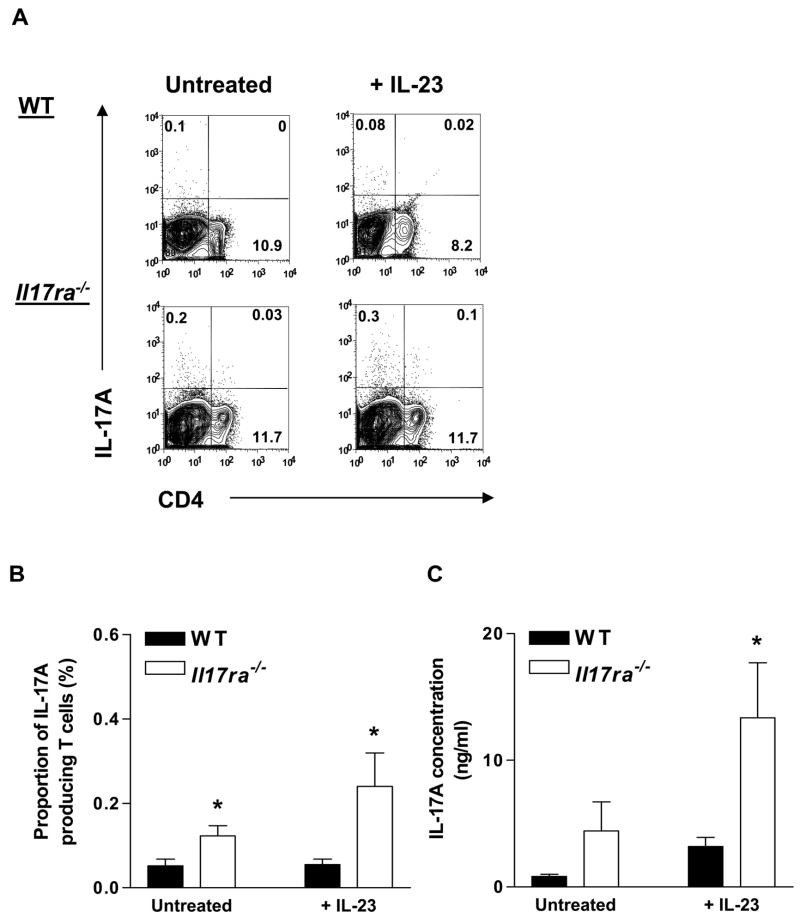
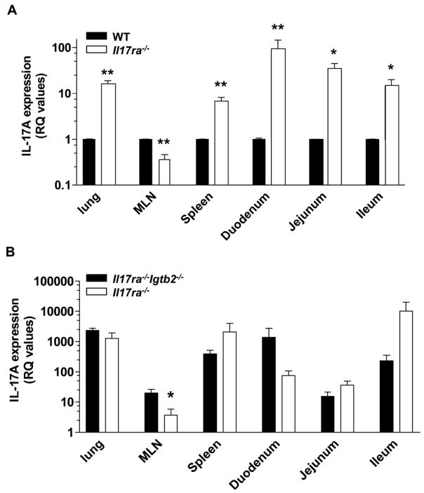
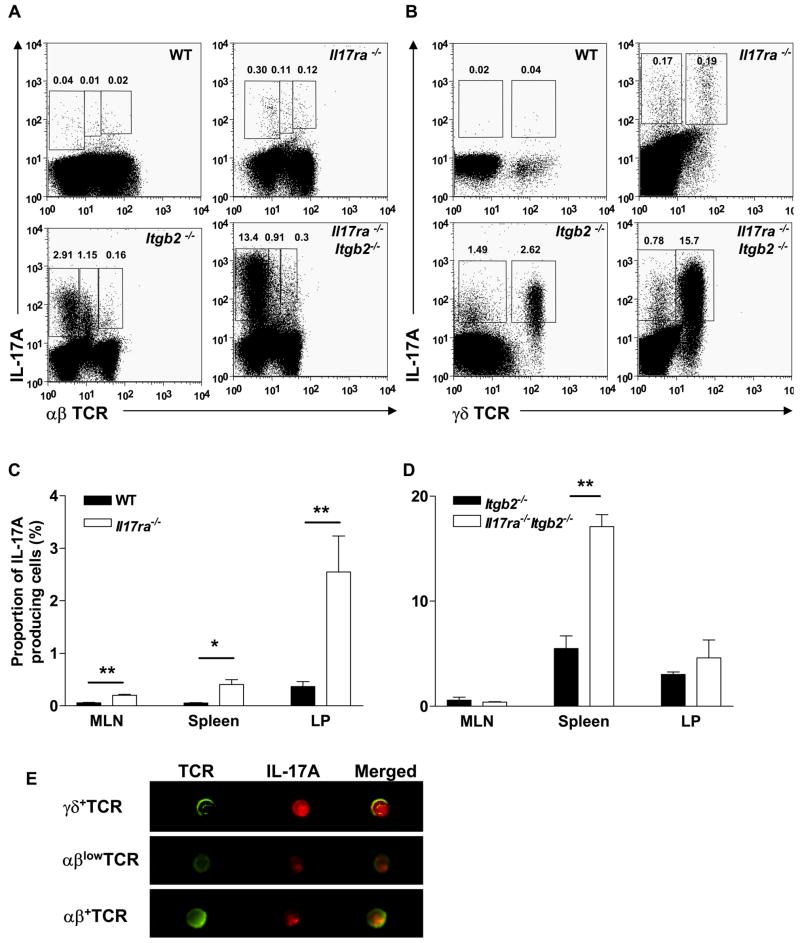
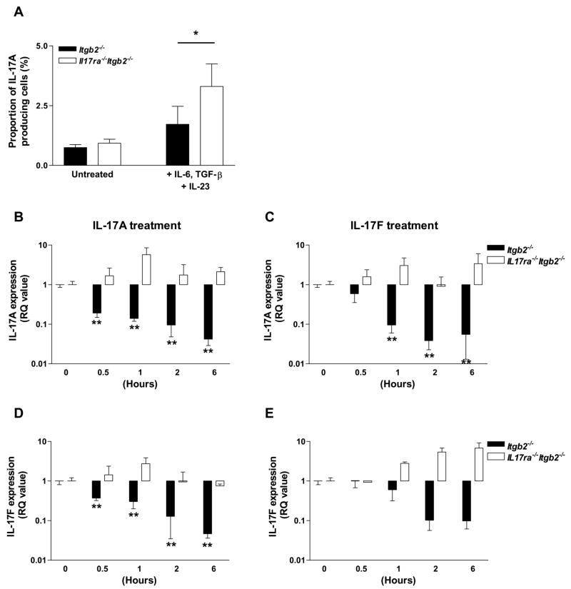
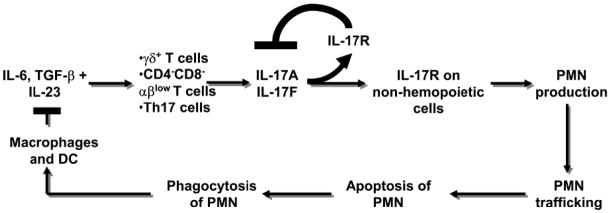
IL-23 and IL-17A regulate granulopoiesis through G-CSF, the main granulopoietic cytokine. IL-23 is secreted by activated macrophages and dendritic cells and promotes the expansion of three subsets of IL-17A-expressing neutrophil-regulatory T (Tn) cells; CD4(-)CD8(-)alphabeta(low), CD4(+)CD8(-)alphabeta(+) (Th17), and gammadelta(+) T cells. In this study, we investigate the effects of IL-17A on circulating neutrophil levels using IL-17R-deficient (Il17ra(-/-)) mice and Il17ra(-/-)Itgb2(-/-) mice that lack both IL-17R and all four beta(2) integrins. IL-17R deficiency conferred a reduction in neutrophil numbers and G-CSF levels, as did Ab blockade against IL-17A in wild-type mice. Bone marrow transplantation revealed that IL-17R expression on nonhemopoietic cells had the greatest effects on regulating blood neutrophil counts. Although circulating neutrophil numbers were reduced, IL-17A expression, secretion, and the number of IL-17A-producing Tn cells were elevated in Il17ra(-/-) and Il17ra(-/-)Itgb2(-/-) mice, suggesting a negative feedback effect through IL-17R. The negative regulation of IL-17A-producing T cells and IL-17A and IL-17F gene expression through the interactions of IL-17A or IL-17F with IL-17R was confirmed in splenocyte cultures in vitro. We conclude that IL-17A regulates blood neutrophil counts by inducing G-CSF production mainly in nonhemopoietic cells. IL-17A controls the expansion of IL-17A-producing Tn cell populations through IL-17R.


Figures

Figure 1
Figure 1. Blood neutrophil counts are reduced in IL-17R-deficient mice
Blood neutrophil counts were measured in (A) IL-17R-deficient mice (n=14) compared to WT mice (n=15), in (B) Il17ra−/−Itgb2−/− mice (n=7) compared to Itgb2−/− mice (n=22) and in (C) WT mice following antibody treatment against IL-17A for 24h (n=4–8; *p<0.05 by nonparametric Mann-Whitney test). BM transplantations (D) from Il17ra−/−Itgb2−/− or Itgb2−/− donors into irradiated WT or Il17ra−/− recipients (n=5–10) were performed and blood counts assessed after 6 weeks. p<0.05 from all other groups, #p<0.05 compared to Itgb2−/− BM into WT, §p<0.05 compared to Il17ra−/−Itgb2−/− BM into WT mice.
Figure 2
Figure 2. Effects of IL-17R deficiency on serum cytokine levels
Analysis of IL-17A (n=3–6) (A) and G-CSF (n=6) (B) in the serum of WT, Il17ra−/−, Itgb2−/− and Il17ra−/−Itgb2−/− mice. Detection limit of each ELISA assay indicated by dotted line. *p<0.05, **p<0.01 by nonparametric Mann-Whitney test.
Figure 3
Figure 3. Effects of IL-17R deficiency on the number of IL-17A-producing Tn cells and IL-17A secretion
Splenocytes from Il17ra−/− and WT mice were stimulated for 3 days on plate-adsorbed anti-CD3ε and soluble anti-CD28 antibodies in the presence or absence of IL-23 (20 ng/ml), followed by a 3 day rest. Cells were reactivated by PMA/ionomycin in the presence of GolgiStop for 5 h before intracellular staining for IL-17A. (A) Representative plots from WT and Il17ra−/− mice. (B) IL-17A-producing Tn cells (n=3–4) as a proportion (%) of the gated lymphocyte population in Il17ra−/− splenocytes (open bars) compared to WT splenocytes (filled bars). (C) IL-17A protein in the supernatants (n=3–5). *p<0.05 by unpaired t test.
Figure 4
Figure 4. IL-17A is upregulated in the tissue of IL-17R-deficient mice
Basal expression of IL-17A was examined in organs of (A) IL-17R-deficient mice (open bars) compared to WT (filled bars) (n=3–4) and (B) Il17ra−/−Itgb2−/− mice (open bars) compared to Itgb2−/− mice (filled bars) (n=3–5). For the Il17ra−/−, Itgb2−/− and Il17ra−/−Itgb2−/− mice, the relative quantification (RQ) values were expressed as a fold-increase compared to IL-17A expression in WT mice (the calibrator control which is equal to 1). *p<0.05, **p<0.01 by unpaired students t test
Figure 5
Figure 5. IL-17A-producing Tn cell populations are expanded in IL-17R-deficient mice
(A) αβhigh and αβlow T cells and (B) γδ+ T cells in the spleen of WT, IL17R−/−, Itgb2−/−and Il17ra−/−Itgb2−/− mice. IL-17A-producing Tn cell populations are elevated in the MLN, spleen and LP of Il17ra−/− mice compared to WT mice (n=4–12) (C), and in the spleen of Il17ra−/−Itgb2−/− mice compared to Itgb2−/− mice (n=3–13) (D). Representative images of IL-17A-producing Tn cell subsets taken by Amnis (E). *p<0.05, **p<0.01 by unpaired student’s t test.
Figure 6
Figure 6. IL-17A and F negatively regulate their own expression and expansion of IL-17A-producing Tn cells
Unfractionated splenocytes from Itgb2−/− or Il17ra−/−Itgb2−/− mice were cultured on anti-CD3ε and soluble anti-CD28, in the presence or absence of IL-6, TGF-β and IL-23 for 3 days followed by a 3 day rest. Cells were reactivated by PMA/ionomycin in the presence of GolgiStop for 5 h before intracellular staining for IL-17A. Proportion of IL-17A-producing Tn cells (A) in cytokine-stimulated Il17ra−/−Itgb2−/−cultures (open bars) compared to Itgb2−/− cultures (closed bars) (n=3–4). *p<0.05 by unpaired student’s t test. IL-17A (B; p<0.0001 and C; p<0.001 by one-way ANOVA), and IL-17F (D; p<0.0001 and E; p<0.01 by one-way ANOVA) gene expression was determined in unfractionated Itgb2−/− (n=3–4) or Il17ra−/−Itgb2−/− (n=2) splenocyte cultures treated with IL-17A (10 ng/ml) or IL-17F (10 ng/ml) from 0–6h. *p<0.05, **p<0.01 compared to untreated samples (0h) by Bonferroni’s post-hoc test.
Figure 7
Figure 7. Neutrostat feedback loop
Cytokines released by macrophages and dendritic cells (DC) induce the release of IL-17A from three Tn cell subsets. IL-17A binds to IL-17R on non-hemopoietic cells in the bone marrow inducing G-CSF release and elevating blood neutrophil (PMN) counts. PMN traffic into the tissue, undergo apoptosis and become engulfed by tissue resident macrophages and DC. Phagocytosis of apoptotic PMN curbs IL-23 release from these cells, closing the “long” feedback loop.(12,13) The present findings add a “short” feedback loop (bold lines) which negatively regulates IL-17A and IL-17F expression via IL-17R.

References

    1. Kolls JK, Linden A. Interleukin-17 family members and inflammation. Immunity. 2004;21:467–476. - PubMed
    1. Aggarwal S, Ghilardi N, Xie MH, de Sauvage FJ, Gurney AL. Interleukin-23 promotes a distinct CD4 T cell activation state characterized by the production of interleukin-17. J Biol Chem. 2003;278:1910–1914. - PubMed
    1. Nakae S, Saijo S, Horai R, Sudo K, Mori S, Iwakura Y. IL-17 production from activated T cells is required for the spontaneous development of destructive arthritis in mice deficient in IL-1 receptor antagonist. PNAS. 2003;100:5986–5990. - PMC - PubMed
    1. Nakae S, Nambu A, Sudo K, Iwakura Y. Suppression of Immune Induction of Collagen-Induced Arthritis in IL-17-Deficient Mice. J Immunol. 2003;171:6173–6177. - PubMed
    1. Miossec P. IL-17 in rheumatoid arthritis: a new target for treatment or just another cytokine? Joint Bone Spine. 2004;71:87–90. - PubMed

Publication types

MeSH terms