. 2008 Sep 12;283(37):25455-25467.
doi: 10.1074/jbc.M801934200. Epub 2008 Jul 7.

Hepatitis B virus X protein via the p38MAPK pathway induces E2F1 release and ATR kinase activation mediating p53 apoptosis

Affiliations

Hepatitis B virus X protein via the p38MAPK pathway induces E2F1 release and ATR kinase activation mediating p53 apoptosis

Wen-Horng Wang et al. J Biol Chem. .

Abstract

Hepatitis B virus (HBV) X protein (pX) is implicated in hepatocellular carcinoma (HCC) pathogenesis by an unknown mechanism. Deletions or mutations of genes involved in the p53 pathway are often associated with HBV-mediated HCC, indicating rescue from p53 apoptosis is a likely mechanism in HBV-HCC pathogenesis. Herein, we determined the mechanism by which pX sensitizes hepatocytes to p53-mediated apoptosis. Although it is well established that the Rb/E2F/ARF pathway stabilizes p53, and the DNA damage-activated ATM/ATR kinases activate p53, the mechanism that coordinates these two pathways has not been determined. We demonstrate that the p38MAPK pathway activated by pX serves this role in p53 apoptosis. Specifically, the activated p38MAPK pathway stabilizes p53 via E2F1-mediated ARF expression, and also activates the transcriptional function of p53 by activating ATR. Knockdown of p53, E2F1, ATR, or p38MAPKalpha abrogates pX-mediated apoptosis, demonstrating that E2F1, ATR, and p38MAPKalpha are all essential in p53 apoptosis in response to pX. Specifically, in response to pX expression, the p38MAPK pathway activates Cdk4 and Cdk2, leading to phosphorylation of Rb, release of E2F1, and transcription of ARF. The p38MAPK pathway also activates ATR, leading to phosphorylation of p53 on Ser-18 and Ser-23, transcription of pro-apoptotic genes Bax, Fas, and Noxa, and apoptosis. In conclusion, pX sensitizes hepatocytes to p53 apoptosis via activation of the p38MAPK pathway, which couples p53 stabilization and p53 activation, by E2F1 induction and ATR activation, respectively.


Figures
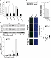
FIGURE 1.
FIGURE 1.
pX expression mediates apoptosis via p53. A, transient transfections of p53-Luc (100 ng), pBax-Luc (100 ng), pFas-Luc (100 ng), pNFκB-Luc (100 ng), and NFAT-Luc (100 ng) plasmids in apoptotic 4pX-1 cells grown as a function of pX expression, with (+) or without (-) PFT-α (10 μm), as indicated. pX expression is via the Tet-off system, by tetracycline removal (5 μg/ml). Results are expressed as pX-dependent induction, -Tet/+Tet ratio, quantified from three independent assays performed in triplicate. B, immunoblot of Bax and p21 employing WCE from apoptotic 4pX-1 cultures grown with (+) or without (-) pX. Actin is the internal control. Quantification is by the Scion software, relative to actin. C, pX-dependent apoptosis, -Tet/+Tet ratio, by PhosphoImager quantification of three independent radioactive DNA fragmentation assays of apoptotic 4pX-1 cultures (21) grown with (+) or without (-) PFT-α, as indicated. D, upper left panel, Western blot of p53 using WCE from 4pX-1 and 4pX-1-p53kd cell lines grown with (+) pX in apoptotic conditions for 2 h. Upper right panel, transient transfections of p53-Luc (100 ng) and pBax-Luc (100 ng) plasmids in 4pX-1-p53kd cells, grown as a function of pX and expressed as the -Tet/+Tet ratio. Lower left panel, immunofluorescence microscopy using the fluorigenic Z-DEVD-FMK caspase3 substrate (21) and 4pX-1 and 4pX-1-p53kd cells grown in apoptotic conditions for 24 h with (+) or without (-) pX or PFT-α (10 μm), as indicated. Lower right panel shows flow cytometric quantification of caspase3-positive cells in 4pX-1 and 4pX-1-p53kd cell lines, using the fluorigenic Z-DEVD-FMK caspase3 substrate (21). Error bars in A, C, and D represent the S.D.
FIGURE 2.
FIGURE 2.
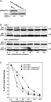
p53 stabilization by pX via the p38MAPK pathway. A, Western blot of p38MAPKα using WCE from 4pX-1 and 4pX-1-p38MAPKαkd cell lines. Quantification is by the Scion software, relative to actin. B, confluent 4pX-1 and 4pX-1-p38MAPKαkd cells grown with (+) or without (-) pX, were incubated in 2% fetal calf serum to initiate apoptosis (21). Cycloheximide (10 μm) was added 2 h after onset of apoptosis, for the indicated time course. WCE isolated 0–180 min after treatment with cycloheximide, immunoblotted for p53. Actin is the internal control. A representative assay is shown from at least three independent experiments. C, quantification by the Scion software of p53 immunoblots shown in B, expressed as % p53 remaining versus min of cycloheximide treatment. Quantification is relative to actin, from at least three independent experiments. Error bars represent S.D.
FIGURE 3.
FIGURE 3.
pX-mediated p38MAPK activation induces Cdk4/Cdk2 leading Rb phosphorylation. A, transient transfections of E2F1-responsive plasmids APAF-Luc (100 ng) and CyclA-Luc (100 ng) in apoptotic 4pX-1 cells grown as a function of pX expression. Results are expressed as pX-dependent induction, -Tet/+Tet ratio, quantified from three independent experiments performed in triplicate. Error bars are S.D. B, Western blot assays using WCE from apoptotic 4pX-1 and 4pX-1-p38MAPKαkd cells grown with (+) or without (-) pX as indicated, employing antibodies specific for phospho-Ser800/804-Rb and Rb. Actin is the loading control. Quantification performed by the Scion software, is expressed as relative intensity to total Rb. A representative assay is shown from three independent experiments. C, upper panel, in vitro Cdk4 immunocomplex kinase assays of WCE (2 mg) from apoptotic 4pX-1 cells grown with (+) or without (-) pX, SB202190 (5 μm) or SP600125 (5 μm), as indicated, using GST-Rb as substrate and [γ-32P]ATP. Reactions were analyzed by SDS-PAGE and autoradiography. pX-dependent induction quantified by PhosphorImager is the -Tet/Tet ratio. Lower panel, Western blot analyses of Cdk4 immunoprecipitates with the Cdk4 antibody. D, Western blot analyses of Cdk2 immunoprecipitates using the phospho-Tyr15-Cdc2 antibody. WCE were isolated from apoptotic 4pX-1 and 4pX-1-p38MAPKαkd cells grown with (+) or without (-) pX, at 0 and 12 h after onset of apoptosis. Quantification is relative to Cdk2 (lower panel), using the Scion software.
FIGURE 4.
FIGURE 4.
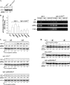
pX induces E2F1 release and ARF expression. A, Western blot of E2F1 using WCE from 4pX-1 and 4pX-1-E2F1kd cell lines grown with (+)pXin apoptotic conditions for 2 h. Quantification is by the Scion software, relative to actin. B, ChIP employing E2F1-specific antibody and apoptotic 4pX-1 and 4pX-1-E2F1kd cells grown with (+) or without (-) pX, with PCR primers for the E2F1 binding sites of ARF, Chk2, and ASPP2 promoters. Left panel, quantification of ChIP assays by real-time PCR from three independent assays. Relative to IgG the pX-dependent induction is significant (p < 0.005). Right panel, agarose gel electrophoresis of PCR products from ChIP assays immunoprecipitated with E2F1 antibody or IgG using apoptotic 4pX-1 cells grown with (+) or without (-) pX. C, Western blot of ARF using WCE from apoptotic 4pX-1, 4pX-E2F1kd, and 4pX-1-p38MAPKαkd cell lines grown with (+) or without (-) pX, as indicated. Actin is the internal control. Quantification relative to actin is by the Scion software. D, Western blot of p53 using WCE isolated from apoptotic 4pX-1, 4pX-E2F1kd, and p38MAPKαkd cell lines grown with (+) or without (-) pX, as indicated. Quantification relative to actin is by the Scion software.
FIGURE 5.
FIGURE 5.
pX expression in apoptotic conditions by growth factor withdrawal activates ATR but not ATM. A, Western blot analyses using WCE from apoptotic 4pX-1 cells grown with (+) or without (-) pX and caffeine (8 mm) as indicated, with antibodies specific for phospho-ATR, ATR, phosphor-Ser1987-ATM, ATM, and p53. Actin is the loading control. Control lanes include treatment of 4pX-1 cells with UV for ATR activation, or with 0.5 μg/ml doxorubicin for ATM activation. B, Western blot of ATR using WCE isolated from 4pX-1 and 4pX-ATRkd cell lines. C, Western blot analyses of p53 using WCE isolated from apoptotic 4pX-1 and 4pX-ATRkd cell lines grown with (+) or without (-) pX, as indicated. Quantification (A–C) is by the Scion software. A representative assay is shown from three independent experiments.
FIGURE 6.
FIGURE 6.
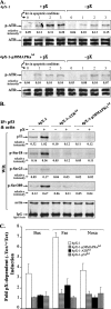
pX activates ATR via the p38MAPK pathway leading to p53 transcriptional activation. A, Western blot analyses of phospho-ATR using WCE from apoptotic 4pX-1 and 4pX-1-p38MAPKαkd cells grown with (+) or without (-) pX, as indicated. ATR is the internal control. B, WCE (2 mg) from 4pX-1, 4pX-1-ATRkd, and 4pX-1-p38MAPKαkd cells grown with (+) or without (-) pX, isolated at 2 h following the onset of apoptosis, were immunoprecipitated with p53 and actin antibodies added in the same reaction. Immunoprecipitates were analyzed by Western blots using antibodies for p53, phospho-Ser18, phospho-Ser23, and phospho-Ser389 of p53. Actin and IgG are used as immunoprecipitation controls. Quantification for A and B is by the Scion software. A representative assay is shown from three independent experiments. C, real-time PCR quantification of Bax, Fas, and Noxa mRNAs employing RNA isolated from 4pX-1, 4pX-ATRkd, and 4pX-1-p38MAPKαkd cell lines grown in apoptotic conditions for 12 h as a function of pX. Results, expressed as pX-dependent induction, -Tet/+Tet ratio, are from three independent RNA isolations, each PCR reaction performed in identical triplicates. Quantification is relative to 18 S rRNA. The pX-dependent induction of Bax, Fas, and Noxa mRNAs in 4pX-1 cells relative to the knockdown cell lines is significant (p < 0.005).
FIGURE 7.
FIGURE 7.
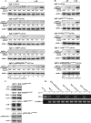
E2F1, ATR and p38MAPKα are essential in p53 apoptosis in response to pX. A and B, Western blot assays of active caspase3 using WCE isolated from (A) apoptotic 4pX-1 and 4pX-1-p53kd, 4pX-1-E2F1kd, 4pX-1-ATRkd, and 4pX-1-p38MAPKαkd cells or (B) the respective pseudo-knockdown cell lines, grown with (+) or without (-) pX as indicated. Quantification is by the Scion software. A representative assay is shown from three independent experiments. C, Western blot assays of p53, E2F1, ATR, and p38MAKα using WCE from 4pX-1 cells and the indicated pseudo-knockdown cell lines. D, agarose gel electrophoresis of PCR reactions employing RNA isolated from the indicated cell lines grown with (+) or without (-) tetracycline for 24 h. pT7-X is the positive control. GAPDH is the internal control.
FIGURE 8.
FIGURE 8.
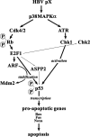
Mechanism of p53 activation by pX. pX stabilizes p53 by activating p38MAPK, leading to Cdk4- and Cdk2-mediated E2F1 release and ARF expression. E2F1 also induces transcription of additional E2F1 genes, including ASPP2 and Chk2. The pX-mediated p38MAPK activation induces ATR activation, leading to p53 activation, p53 pro-apoptotic gene transcription, and apoptosis. Furthermore, ATR activation contributes to p53 stabilization upon serum withdrawal as shown in Fig. 6B.

References

    1. Kinzler, K. W., and Vogelstein, B. (1997) Nature 386 761-763 - PubMed
    1. Giaccia, A. J., and Kastan, M. B. (1998) Genes Dev. 12 2973-2983 - PubMed
    1. Lowe, S. W., and Ruley, H. T. (1993) Genes Dev. 7 535-545 - PubMed
    1. de Stanchina, E., McCurrach, M. E., Zindy, F., Shieh, S.-Y., Ferbeyer, G., Samuelson, A. V., Prives, C., Roussel, M. F., Sherr, C. J., and Lowe, S. (1998) Genes Dev. 12 2434-2442 - PMC - PubMed
    1. Ginsberg, D. (2002) FEBS Lett. 529 122-125 - PubMed

Publication types

MeSH terms

LinkOut - more resources