. 2008 Sep 5;283(36):25074-81.
doi: 10.1074/jbc.M801214200. Epub 2008 Jul 7.

High density lipoprotein-associated sphingosine 1-phosphate promotes endothelial barrier function

Affiliations

High density lipoprotein-associated sphingosine 1-phosphate promotes endothelial barrier function

Kelley M Argraves et al. J Biol Chem. .

Abstract

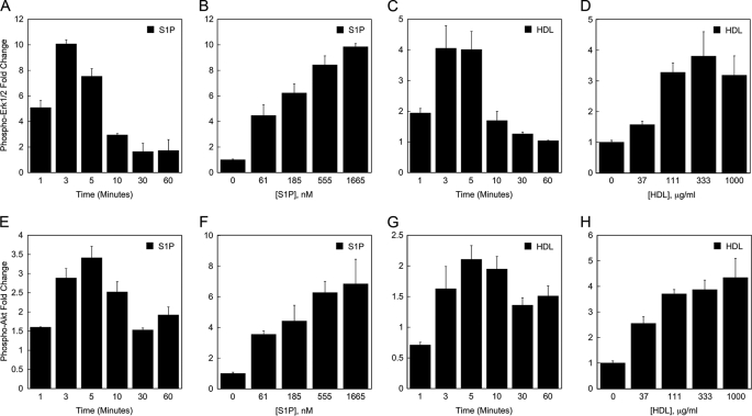
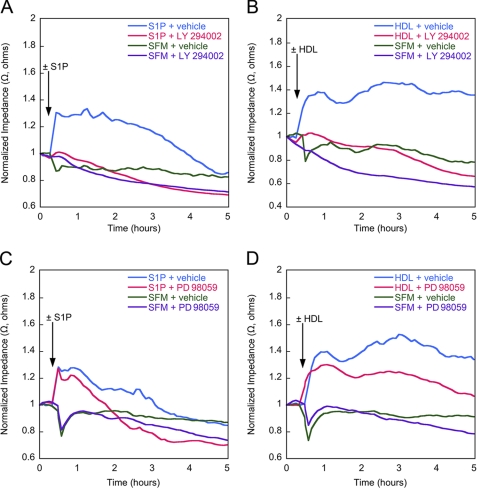
High density lipoproteins (HDL) are major plasma carriers of sphingosine 1-phosphate (S1P). Here we show that HDL increases endothelial barrier integrity as measured by electric cell substrate impedance sensing. S1P was implicated as the mediator in this process through findings showing that pertussis toxin, an inhibitor of Gi-coupled S1P receptors, as well as antagonists of the S1P receptor, S1P1, inhibited barrier enhancement by HDL. Additional findings show that HDL stimulates endothelial cell activation of Erk1/2 and Akt, signaling pathway intermediates that have been implicated in S1P-dependent endothelial barrier activity. HDL was also found to promote endothelial cell motility, a process that may also relate to endothelial barrier function in the context of a vascular injury response. The effects of HDL on endothelial cell Erk1/2 and Akt activation and motility were suppressed by pertussis toxin and S1P1 antagonists. However, both HDL-induced barrier enhancement and HDL-induced motility showed a greater dependence on Akt activation as compared with Erk1/2 activation. Together, the findings indicate that HDL has endothelial barrier promoting activities, which are attributable to its S1P component and signaling through the S1P1/Akt pathway.


Figures

FIGURE 1.
FIGURE 1.
HDL enhances transendothelial electrical resistance. A minimal TEER plateau was reached within ∼24 h of replacing the culture medium of confluent endothelial cells with serum-free medium. The monolayers were then incubated with varying concentrations S1P (A) or HDL (B). In B, the concentration of S1P in the HDL tested ranged from 5 to 403 nm. The TEER tracings shown in panels A and B each represent mean data from three independent experiments each with two replicates per condition. As a control, monolayers were treated with 40 μg/ml BSA, a concentration corresponding to the amount of BSA carrier used for the highest concentration of S1P tested. Impedance values were normalized by dividing each value by the level of impedance measured just prior to the addition of effectors.
FIGURE 2.
FIGURE 2.
HDL-enhanced transendothelial electrical resistance is inhibited by pertussis toxin and S1P1 antagonists. Confluent endothelial monolayers were grown under serum-free conditions until a minimal TEER plateau had been reached. The cells were then incubated with S1P or HDL in the presence or absence of PTX (A and B) or S1P1 antagonists (C and D). In A and B, S1P was used at 833 nm, HDL was used at 1000 μg/ml (containing 400 nm S1P), and PTX was used at 1 μg/ml. In C and D, S1P was used at 250 nm, HDL was used at 621 μg/ml (containing 250 nm S1P), the S1P1 antagonist 857390 was used at 10 μm, and the S1P1/S1P3 antagonist VPC23019 was used at 10 μm. As controls, monolayers were treated with BSA-containing serum-free medium (SFM) plus or minus vehicle buffer. Each of the TEER tracings shown is an average of two replicate wells and representative of three independent experiments. Impedance values were normalized by dividing each value by the level of impedance measured just prior to the addition of effectors.
FIGURE 3.
FIGURE 3.
HDL stimulates Erk1/2 and Akt activation in endothelial cells. The effect of S1P and HDL on activation of Erk1/2 (A-D) and Akt (E-H) in HUVEC was determined by multiplex bead array assay. In A-D, the values for the -fold difference in Erk1/2 phosphorylation were derived from the level of phospho-Erk1/2 fluorescence in S1P- or HDL-treated cells divided by the level of phospho-Erk1/2 fluorescence in control cells. In E-G, the values for the -fold increase in Akt phosphorylation were derived from the level of phospho-Akt fluorescence in cells treated with S1P or HDL divided by the level of phospho-Akt fluorescence measured in control cells. The data depicted in panels A, C, E, and G is based on treating HUVEC for the indicated times with 833 nm S1P or 333 μg/ml HDL (containing 133 nm S1P). The data depicted in panels B, D, F, and H are based on treating HUVEC with the indicated concentrations of S1P or HDL for 3 min. The level of S1P in the 3-fold dilutions of HDL tested in panel H ranged from 12 to 337 nm. Data are shown from a representative experiment. Each experiment was performed two times; each data point is an average from two independent wells.
FIGURE 4.
FIGURE 4.
HDL activation of Erk1/2 and Akt in HUVEC is inhibited by pertussis toxin and S1P1 antagonists. In A and B, PTX (100 ng/ml) or the PTX buffer was added to EBM during the final 12 h of serum starvation. BSA, S1P (200 nm), or HDL (containing 200 nm S1P) were then added to the medium and allowed to incubate with the cells for 3 min. In C and D, the S1P1 antagonist 857390 or the S1P1/S1P3 antagonist or vehicle was added to the medium 15 min prior to addition of S1P or HDL and allowed to incubate with the cells for 3 min. The graphed values were derived from the level of phospho-Erk1/2 or phospho-Akt fluorescence in S1P- or HDL-treated cells divided by the level of phospho-Erk1/2 or phospho-Akt fluorescence in BSA-treated cells. Data are shown from representative experiments, antagonist and inhibitor experiments were performed 2-4 times (e.g. PTX, n = 3; S1P1 antagonist, n = 4; S1P1/S1P3 antagonist, n = 2). Each data point is an average from two independent wells.
FIGURE 5.
FIGURE 5.
Inhibition of Akt and Erk1/2 blocks HDL-induced enhancement of TEER. Confluent endothelial monolayers were grown under serum-free conditions until a minimal TEER plateau had been reached. The cells were then incubated with S1P or HDL in the presence or absence of the Akt inhibitor LY294002 ((LY) A and B) or the Erk inhibitor PD98059 ((PD) C and D). Each of the TEER tracings shown is an average of two replicate wells and representative of three independent experiments. Impedance values were normalized by dividing each value by the level of impedance measured just prior to the addition of effectors. SFM, serum-free medium.
FIGURE 6.
FIGURE 6.
HDL promotes S1P receptor-dependent endothelial migration in ECIS wounding assay. HUVEC grown on the microelectrodes of the ECIS wells were killed with a burst of high electrical current. Culture medium was supplemented with S1P (833 nm) or HDL (1000 μg/ml containing 400 nm S1P) in the presence or absence of PTX (A and B) or S1P1 antagonists (C and D), and the migration of cells into the wound areas was measured in real time by electrical impedance. Data depicted in each of the TEER tracings are the average of two independent experiments each with two replicates per condition. SFM, serum-free medium.
FIGURE 7.
FIGURE 7.
HDL-stimulated endothelial migration is dependent on Akt activation to greater extent than Erk activation. HUVEC grown on the microelectrodes of the ECIS wells were killed with a burst of high electrical current. One hour before the electrical wounding, the culture medium was supplemented with the Akt inhibitor LY294002 ((LY) A and B) or the Erk inhibitor PD98059 ((PD) C and D). S1P (250 nm) and HDL (234 μg/ml containing 250 nm S1P) were added after electrical wounding. Each of the TEER tracings shown is representative of three independent experiments. SFM, serum-free medium.

References

    1. Garcia, J. G., Liu, F., Verin, A. D., Birukova, A., Dechert, M. A., Gerthoffer, W. T., Bamberg, J. R., and English, D. (2001) J. Clin. Investig. 108689 -701 - PMC - PubMed
    1. Schaphorst, K. L., Chiang, E., Jacobs, K. N., Zaiman, A., Natarajan, V., Wigley, F., and Garcia, J. G. (2003) Am. J. Physiol. 285L258 -L267 - PubMed
    1. Xu, M., Waters, C. L., Hu, C., Wysolmerski, R. B., Vincent, P. A., and Minnear, F. L. (2007) Am. J. Physiol. 4C1309 -C1318 - PubMed
    1. Peng, X., Hassoun, P. M., Sammani, S., McVerry, B. J., Burne, M. J., Rabb, H., Pearse, D., Tuder, R. M., and Garcia, J. G. (2004) Am. J. Respir. Crit. Care Med. 1691245 -1251 - PubMed
    1. Lee, J. F., Zeng, Q., Ozaki, H., Wang, L., Hand, A. R., Hla, T., Wang, E., and Lee, M. J. (2006) J. Biol. Chem. 28129190 -29200 - PubMed

Publication types

MeSH terms