. 2008 Jul 14;182(1):141-55.
doi: 10.1083/jcb.200712094. Epub 2008 Jul 7.

SUMOylation of the MAGUK protein CASK regulates dendritic spinogenesis

Affiliations

SUMOylation of the MAGUK protein CASK regulates dendritic spinogenesis

Hsu-Wen Chao et al. J Cell Biol. .

Abstract

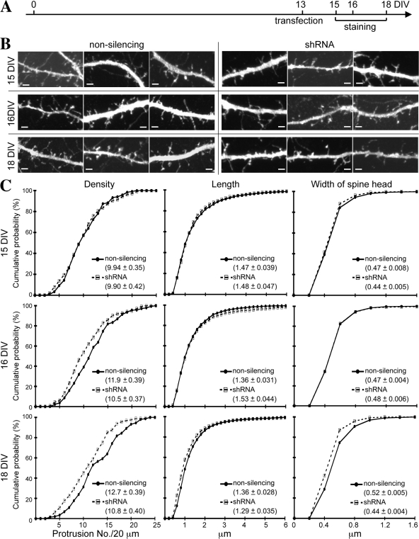
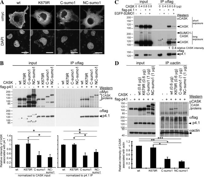
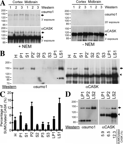
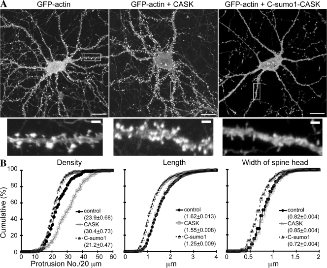
Membrane-associated guanylate kinase (MAGUK) proteins interact with several synaptogenesis-triggering adhesion molecules. However, direct evidence for the involvement of MAGUK proteins in synapse formation is lacking. In this study, we investigate the function of calcium/calmodulin-dependent serine protein kinase (CASK), a MAGUK protein, in dendritic spine formation by RNA interference. Knockdown of CASK in cultured hippocampal neurons reduces spine density and shrinks dendritic spines. Our analysis of the time course of RNA interference and CASK overexpression experiments further suggests that CASK stabilizes or maintains spine morphology. Experiments using only the CASK PDZ domain or a mutant lacking the protein 4.1-binding site indicate an involvement of CASK in linking transmembrane adhesion molecules and the actin cytoskeleton. We also find that CASK is SUMOylated. Conjugation of small ubiquitin-like modifier 1 (SUMO1) to CASK reduces the interaction between CASK and protein 4.1. Overexpression of a CASK-SUMO1 fusion construct, which mimicks CASK SUMOylation, impairs spine formation. Our study suggests that CASK contributes to spinogenesis and that this is controlled by SUMOylation.


Figures

Figure 1.
Figure 1.
Knockdown of endogenous CASK prevents dendritic spine formation in cultured hippocampal neurons. (A) CASK shRNA reduced the expression of endogenous CASK in HEK cells. The CASK shRNA construct was transfected into HEK cells. 1 d later, cells were harvested for immunoblotting analysis using CASK and tubulin antibodies. Nonsilencing construct was used as a negative control. Tubulin was used as an internal control. (B) CASK shRNA down-regulated overexpressed Myc-tagged rat CASK but not Myc-tagged PSD-95, Myc-tagged SAP-97, or a Myc-tagged rat CASK silent mutant insensitive to CASK shRNA. COS cells were cotransfected with CASK shRNA or nonsilencing construct and various MAGUK protein expression plasmids as indicated and were harvested 1 d later for immunoblotting using Myc tag antibody. Tubulin was used as an internal control. (C) Immunostaining of cultured hippocampal neurons showing the reduction of endogenous CASK by CASK shRNA. Cultured hippocampal neurons were cotransfected with CASK shRNA construct and GFP-actin at 14 DIV. 2 d later, cells were stained with CASK antibody. Two representative images are shown. Arrowheads point to the transfected neurons, which are GFP-actin positive. Higher magnifications of the boxed areas in the middle panel are shown on the right. More images showing the knockdown effect of CASK shRNA are included in Fig. S1 (available at http://www.jcb.org/cgi/content/full/jcb.200712094/DC1). (D) CASK shRNA prevented dendritic spine formation. Cultured hippocampal neurons were cotransfected with GFP-actin and CASK shRNA, nonsilencing control, or CASK mutant resistant to CASK shRNA as indicated at 13–14 DIV. 5 d later, cells were harvested for fluorescence immunostaining using CASK (visualized by AlexaFluor594) and GFP (visualized by AlexaFluor488) antibodies. Only the GFP patterns are shown. (E) Quantification of the effect of CASK shRNA on dendritic spine morphology. For each experiment, at least 11 transfected neurons were analyzed from two independent experiments. A total of 1,027 (nonsilencing), 798 (CASK shRNA), and 549 (CASK shRNA + CASK mutant) spines were analyzed. Mean values ± SEM for each group are shown. Bars: (C) 20 μm; (D) 1 μm.
Figure 2.
Figure 2.
Time course of CASK knockdown changes in spine morphology. (A) CASK shRNA and nonsilencing control were cotransfected with GFP into hippocampal neurons at 13 DIV. Immunostaining using CASK and GFP antibodies were performed at 15, 16, and 18 DIV as indicated. (B) Representative images of neurons transfected with CASK shRNA and nonsilencing control. Only the GFP signals are shown to outline the morphology of dendrites. The effect of CASK shRNA on down-regulation of CASK protein levels is shown in Fig. S2 (available at http://www.jcb.org/cgi/content/full/jcb.200712094/DC1). (C) Quantification analysis. More than 14 neurons collected from two independent experiments were analyzed for each group. The total number of spines analyzed in each group was as follows: 15 DIV, 1,225 nonsilencing and 1,028 CASK shRNA; 16 DIV, 1,660 nonsilencing and 1,420 CASK shRNA; and 18 DIV, 1,878 nonsilencing and 1,730 CASK shRNA. Mean values ± SEM for each group are shown. Bars, 2 μm.
Figure 3.
Figure 3.
The PDZ domain and protein 4.1–binding site of CASK are involved in dendritic spine formation. (A) Overexpression of CASK PDZ domain (PDZ), the C-terminal tail of syndecan-2 (synd-2C), and the CASKΔp4.1 mutant prevented spinogenesis. Cultured hippocampal neurons were cotransfected with GFP and various constructs as indicated at 13 DIV. 5 d later, cells were harvested for immunostaining using HA tag and GFP antibodies. Only the GFP patterns are shown. (B) Quantitative analysis of A. The total number of spines analyzed in each group was 756 GW1 control, 385 PDZ, 535 Synd-2C, and 494 CASKΔp4.1. More than 10 neurons for each group were analyzed. Mean ± SEM for each group is shown. (C) Overexpression of the CASK PDZ domain and CASKΔp4.1 mutant does not rescue the phenotype of CASK knockdown. At 12 DIV, cultured hippocampal neurons were cotransfected with GFP-actin and various constructs as indicated. Immunostaining using CASK, HA tag, and GFP antibodies was performed at 18 DIV. Only GFP patterns are shown. (D) Quantitative analysis of C. The total number of neurons and spines analyzed in each group was as follows: nonsilencing + GW1 vector, 31 and 2,703; nonsilencing + CASK, 27 and 4,037; shRNA + GW1 vector, 31 and 1,754; shRNA + PDZ, 32 and 1,317; and shRNA + Δp4.1, 31 and 2,085. Mean ± SEM for each group is shown. Bars, 2 μm.
Figure 4.
Figure 4.
CASK is modified by SUMO1 at residue K679. (A) A schematic diagram of CASK, CASK deletion mutants, and SUMO1-CASK fusions. Some of the constructs had two versions: one was Myc tagged at the N-terminal end, and the other had no extra tag. (B) COS1 cells were cotransfected with plasmids expressing 1 μg Myc-tagged SUMO1, 2, or 3 and 1 μg CASK. After 24-h incubation, total cell extracts were harvested and immunoprecipitated with CASK antibody. Immunoblotting analysis was then performed sequentially using CASK antibody to monitor CASK proteins and Myc tag antibody to detect Myc-tagged SUMOs. (C) 1.2 μg CASK deletion mutants was cotransfected with 0.4 μg SUMO1 into COS1 cells to evaluate the SUMO1-modified region of CASK by immunoprecipitation-immunoblotting analysis as described in B except that Myc tag antibody was used first for immunoblotting. The arrowhead points to the position of endogenous full-length CASK in COS1 cells. Arrows indicate the positions of SUMO1-CASK deletion mutants. (D) Two putative SUMO1-modified consensus motifs, guanylate kinase648LE and TK679QE, were predicted in the sequence of rat CASK, and site-directed mutagenesis was performed to change lysine 648 and 679 to arginine. COS1 cells were cotransfected with 0.5 μg SUMO1 and 0.5 μg of wild-type CASK, K648R, or K679R mutant and subjected to immunoprecipitation-immunoblotting analysis. (B and D) Arrows indicate the positions of SUMOylated CASK; arrowheads point to the positions of unmodified full-length CASK.
Figure 5.
Figure 5.
SUMOylation does not promote nuclear distribution of CASK but interferes with the interaction between CASK and protein 4.1N in COS cells. (A) Confocal analysis of the subcellular distribution of CASK proteins. Myc-tagged wild-type and CASK mutants were transfected into COS1 cells as indicated. Nuclei were counterstained with DAPI. (B) The interaction between protein 4.1N and CASK. COS1 cells were cotransfected with 0.25 μg FLAG-tagged protein 4.1N and either 1 μg Myc-tagged CASK or 1 μg CASK mutants as indicated. Immunoprecipitation was performed using FLAG tag antibody. The precipitates were analyzed by immunoblotting using specific antibodies to the Myc and FLAG tags sequentially. The asterisks indicate the residual Myc immunoreactivities from the top panel. The protein levels of coimmunoprecipitated CASK were analyzed by ImageJ version 1.38X (National Institutes of Health). The data presented are the mean ± SEM of three independent experiments. The relative amounts of proteins were normalized to the amounts of CASK input and precipitated protein 4.1 as indicated. The significance of any difference was determined by t test using SPSS software, and significant differences are shown; *, P < 0.05. (C) Overexpression of SUMO1 reduces the interaction between CASK and protein 4.1N. COS cells were triple transfected with CASK, flag-tagged protein 4.1N, and GFP-tagged SUMO1 or vector control and analyzed by immunoprecipitation-immunoblotting using antibodies as indicated. The DNA amounts for transfection are also shown. Because cotransfection with protein 4.1N reduces CASK amounts in the Triton X-100–solubilized lysate (see B), the DNA amounts for transfection were adjusted accordingly to make the CASK protein amounts similar among different groups. The relative protein amounts of CASK coimmunoprecipitated with protein 4.1N were normalized to both precipitated protein 4.1N amounts and CASK input amount. The data are means of two independent experiments. The asterisks indicate the nonspecific signals contributed by precipitated protein 4.1N. (D) SUMOylation reduces the association of CASK with actin cytoskeleton. COS cells were cotransfected with protein 4.1N and CASK constructs as indicated. Immunoprecipitation was performed using actin antibody and analyzed by immunoblotting using antibodies as indicated. The amount of CASK protein coimmunoprecipitated with actin, normalized to actin precipitated amounts, is shown. Data are the means of three independent experiments. Error bars indicate SEM. *, P < 0.05; **, P < 0.005; ***, P < 0.001. Bars, 20 μm.
Figure 6.
Figure 6.
SUMOylated CASK proteins are present in the mouse brain. (A) Mouse brain homogenates purified from three different mice in the presence or absence of N-ethylmaleimide were immunoprecipitated with CASK antibody. The precipitates were then analyzed by immunoblotting using SUMO1 and CASK antibodies sequentially. The exposure time for each image is indicated. (B) Mouse brain subcellular fractions prepared as described in Materials and methods were used to examine the neuronal distribution of SUMOylated CASK. Equal amounts of each fraction were analyzed by immunoprecipitation-immunoblotting analysis as described in A. H, total homogenate; P1, nuclei and cell debris; S1, supernatant of P1; P2′, washed crude synaptosomal fraction; S2, supernatant of P2′; LP1, lysed synaptosomal membrane; LS1, supernatant of LP1; P3, light membrane fraction; S3, soluble cytosol. The asterisk indicates an unknown protein species recognized by SUMO1 antibody. (C) The percentage of SUMOylated CASK in total CASK proteins of each subcellular fraction. The protein levels of SUMOylated CASK and nonSUMOylated CASK were analyzed using ImageJ. For each fraction, the percentage of SUMOylated CASK was determined by dividing the amounts of SUMOylated CASK by the sum of SUMOylated and nonSUMOylated CASK. The data presented are the mean ± SEM (error bars) of four independent experiments. (D) SUMOylated CASK is mostly present in synaptic cytosol, LS2 fraction. The LS1 fraction was further separated into LP2 fraction, crude synaptic vesicles, and LS2 fraction, synaptic cytosol. CASK proteins were immunoprecipitated from equal protein amounts of LP1, LP2, and LS2 fractions and analyzed by immunoblotting using SUMO1 and CASK antibodies as indicated. The percentage of SUMOylated CASK in total CASK proteins is also indicated. (A, B, and D) Arrows indicate the positions of SUMOylated CASK; arrowheads point to the positions of unmodified full-length CASK.
Figure 7.
Figure 7.
Synaptic distribution of SUMOylated CASK. (A) At 16–17 DIV, cultured hippocampal neurons were divided into three different groups: (1) untreated control, (2) 1 μM TTX overnight, and (3) overnight TTX followed by 60 mM KCl for 3 min. 20–40 min after removing KCl, cells were fixed and stained with SUMO1, CASK, and PSD-95 antibodies. (B) Quantification of SUMO1, CASK, and PSD-95 puncta along dendrites. More than 80 dendrites from >22 neurons were analyzed for each group. Mean values ± SEM (error bars) are shown. ***, P < 0.001 by t test. (C) The percentage of PSD-95, CASK, and PSD-95–CASK double-positive puncta in the population of SUMO1-positive puncta along dendrites. (D) Colocalization of PSD-95 and Bassoon along dendrites of neurons treated with TTX alone and TTX followed by potassium chloride. (E) Quantification analysis of PSD-95 and Bassoon double-positive puncta. More than 80 dendrites from >18 neurons were analyzed in each group. Mean values ± SEM are shown. Bars, 2 μm.
Figure 8.
Figure 8.
SUMOylation of CASK influences dendritic spine morphology. (A) Hippocampal neurons were cotransfected with GFP-actin and Myc-CASK, Myc-C-SUMO1-CASK, or vector control at a ratio of 1:5 at 10–12 DIV as indicated and were fixed at 17–18 DIV to examine spine morphology by double staining using Myc and GFP antibodies. Only double-positive neurons were collected for analysis. The images of GFP signal are shown. Myc tag signal revealing the patterns of CASK and C-SUMO1-CASK are shown in Fig. S3 (available at http://www.jcb.org/cgi/content/full/jcb.200712094/DC1). The bottom panel shows higher magnification of the boxed areas in the top panel. (B) Quantification of spine density, length, and width from A. The histograms show the cumulative distribution as a percentage. A total of 2,828 (GFP-actin alone), 5,229 (Myc-CASK), and 3,586 (Myc-C-SUMO1-CASK) spines from >117 dendrites of >22 neurons were analyzed. Mean values ± SEM for each group are shown in the insets. Bars: (A, top) 20 μm; (A, bottom) 2 μm.

References

    1. Alkuraya, F.S., I. Saadi, J.J. Lund, A. Turbe-Doan, C.C. Morton, and R.L. Maas. 2006. SUMO1 haploinsufficiency leads to cleft lip and palate. Science. 313:1751. - PubMed
    1. Atasoy, D., S. Schoch, A. Ho, K.A. Nadasy, X. Liu, W. Zhang, K. Mukherjee, E.D. Nosyreva, R. Fernandez-Chacon, M. Missler, et al. 2007. Deletion of CASK in mice is lethal and impairs synaptic function. Proc. Natl. Acad. Sci. USA. 104:2525–2530. - PMC - PubMed
    1. Biederer, T., and T.C. Sudhof. 2001. CASK and protein 4.1 support F-actin nucleation on neurexins. J. Biol. Chem. 276:47869–47876. - PubMed
    1. Biederer, T., Y. Sara, M. Mozhayeva, D. Atasoy, X. Liu, E.T. Kavalali, and T.C. Sudhof. 2002. SynCAM, a synaptic adhesion molecule that drives synapse assembly. Science. 297:1525–1531. - PubMed
    1. Borg, J.P., S.W. Straight, S.M. Kaech, M. de Taddeo-Borg, D.E. Kroon, D. Karnak, R.S. Turner, S.K. Kim, and B. Margolis. 1998. Identification of an evolutionarily conserved heterotrimeric protein complex involved in protein targeting. J. Biol. Chem. 273:31633–31636. - PubMed

Publication types