. 2008 Aug 4;205(8):1721-8.
doi: 10.1084/jem.20071463. Epub 2008 Jul 7.

Distinct roles of apolipoprotein components within the trypanosome lytic factor complex revealed in a novel transgenic mouse model

Affiliations

Distinct roles of apolipoprotein components within the trypanosome lytic factor complex revealed in a novel transgenic mouse model

Maria Pilar Molina-Portela et al. J Exp Med. .

Abstract

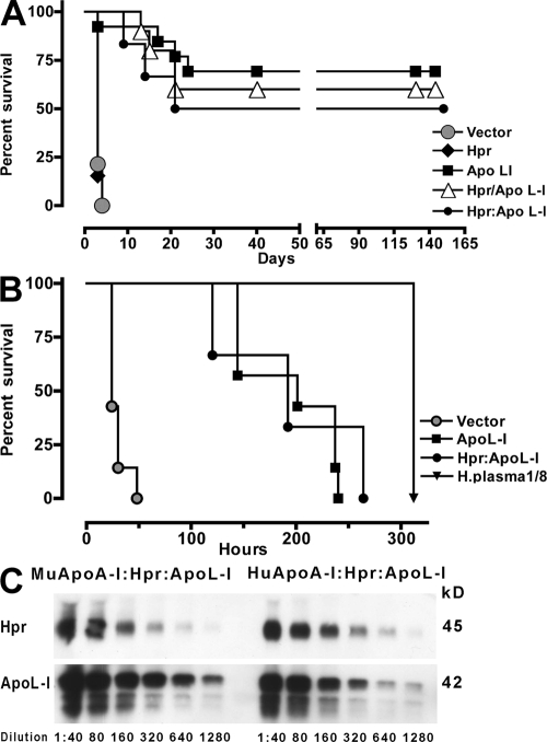
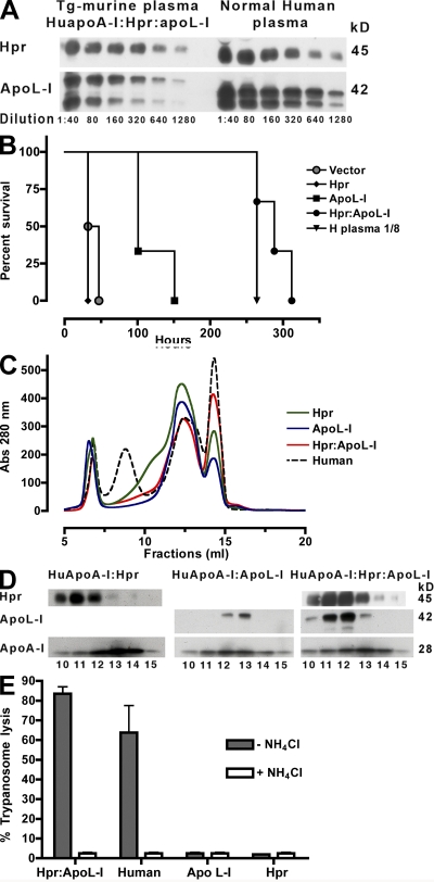
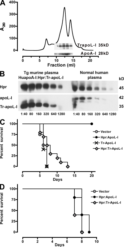
Humans express a unique subset of high-density lipoproteins (HDLs) called trypanosome lytic factors (TLFs) that kill many Trypanosoma parasite species. The proteins apolipoprotein (apo) A-I, apoL-I, and haptoglobin-related protein, which are involved in TLF structure and function, were expressed through the introduction of transgenes in mice to explore their physiological roles in vivo. Transgenic expression of human apolipoprotein L-I alone conferred trypanolytic activity in vivo. Coexpression of human apolipoprotein A-I and haptoglobin-related protein (Hpr) had an effect on the integration of apolipoprotein L-I into HDL, and both proteins were required to increase the specific activity of TLF, which was measurable in vitro. Unexpectedly, truncated apolipoprotein L-I devoid of the serum resistance gene interacting domain, which was previously shown to kill human infective trypanosomes, was not trypanolytic in transgenic mice despite being coexpressed with human apolipoprotein A-I and Hpr and incorporated into HDLs. We conclude that all three human apolipoproteins act cooperatively to achieve maximal killing capacity and that truncated apolipoprotein L-I does not function in transgenic animals.


Figures

Figure 1.
Figure 1.
Survival kinetics of mice infected with T. b. brucei ILTat 1.25, relative lytic units, and protein expression in Tg mice. (A) Mice expressing Hpr (black diamond; n = 13), apoL-I (black square; n = 13), or a combination of Hpr and apoL-I in individual plasmids (white triangle; Hpr/apoL-I; n = 10) or in a single plasmid (black circle, Hpr:apoL-I; n = 6) were infected i.p. with 1.78 × 10⁶ trypanosomes. Control mice were treated with empty vector (gray circle; n = 14). The parasitemia was monitored and time of death was recorded. (B) Survival kinetics of naive mice infected with T. b. brucei ILTat 1.25 that were administered plasma i.v. from mice transfected with empty vector (gray circle; n = 7), apoL-I (black square; n = 7), or a combination of Hpr and apoL-I in a single plasmid (black circle; Hpr:apoL-I; n = 3). The protection obtained by normal human plasma (dilution 1/8) is indicated by the inverted triangle. (C) Western blot with serial dilutions of plasma from Tg mice expressing murine apoA-I and human Hpr and apoL-I from the same plasmid (Hpr:apoL-I) and Tg mice expressing human apoA-I and human Hpr and apoL-I from the same plasmid (Hpr:apoL-I). Hpr and apoL-I were detected with monoclonal antibodies.
Figure 2.
Figure 2.
Increased specific activity of Tg-HDL in the presence of human apoA-I and Hpr. (A) Western blot with serial dilutions of plasma from HuapoA-I mice expressing Hpr and apoL-I from a single plasmid (HuapoA-I:Hpr:apoL-I) and normal human plasma. Hpr and apoL-I were detected with monoclonal antibodies. (B) Survival kinetics of naive mice infected with T. b. brucei ILTat 1.25 and then given 300 μl plasma i.v. from HuapoA-I mice expressing Hpr (black diamond; n = 3), apoL-I (black square; n = 3), or both Hpr and apoL-I from a single plasmid (black circle, Hpr:apoL-I; n = 3). The protection obtained by normal human plasma (dilution 1/8) is indicated by the inverted triangle. (C) A280 profile of KBr-purified lipoproteins from human (dashed line) and HuapoA-I mouse plasma expressing Hpr:apoL-I (red line), Hpr (green line), and apoL-I (blue line) separated by size on a Superdex 200 column; fractions 6–7 are void, fractions 8–9 are human LDL, fractions 10–14 are HDLs, and fraction 15 is albumin. (D) Western blot of different size-fractionated lipoproteins from HuapoA-I mice expressing Hpr only (HuapoA-I:Hpr), apoL-I only (HuapoA-I:apoL-I), or Hpr and apoL-I from the same plasmid (HuapoA-I:Hpr:apoL-I). Hpr and apoL-I were detected with monoclonal antibodies. (E) In vitro lytic activity of size-fractionated HDL (fraction 11) from human and HuapoA-I mice expressing Hpr, apoL-I, or both from a single plasmid (dark gray bars). Open bars represent the lytic activity obtained in the presence of NH4Cl (10 mM). Data show the mean and SEM of five independent measurements.
Figure 3.
Figure 3.
Tr-apoL-I gene expression in Tg-HuapoA-I mice does not protect from infection with T. b. brucei or T. b. brucei-SRA. (A) A280 profile of KBr-purified lipoproteins separated by size on a Superdex 200 column; fractions 6–7 are void, fractions 10–14 are HDLs as indicated by the distribution of apoA-I, and fraction 15 is albumin. (inset) A Western blot of the fractions probed for apoA-I (28 kD) and Tr-apoL-I (∼35 kD) detected with polyclonal anti–apoL-I. (B) Western blot with serial dilutions of plasma from HuapoA-I mice expressing Hpr and Tr-apoL-I from a single plasmid (HuapoA-I:Hpr:Tr-apoL-I) and normal human plasma. Hpr, apoL-I, and Tr-apoL-I were detected with polyclonal antibodies. (C) Mice expressing combination of Hpr and apoL-I in a single plasmid (black circle, Hpr:apoL-I; n = 3), Tr-apoL-I (cross, n = 5), combination of Hpr and Tr-apoL-I in a single plasmid (gray diamond; Hpr:Tr-apoL-I; n = 10) were infected i.p. with 5,000 T. b. brucei trypanosomes. Control mice were treated with empty vector (gray circle; n = 4). The parasitemia was monitored and time of death was recorded. (D) Mice expressing a combination of Hpr and apoL-I in a single plasmid (black circle, Hpr:apoL-I; n = 5) and a combination of Hpr and Tr-apoL-I in a single plasmid (gray diamond; Hpr:Tr-apoL-I; n = 5) were infected i.p. with 5,000 T. b. brucei-SRA trypanosomes. Control mice were treated with empty vector (gray circle; n = 2). The parasitemia was monitored and the time of death was recorded.

References

    1. Ansell, B.J., G.C. Fonarow, and A.M. Fogelman. 2006. High-density lipoprotein: is it always atheroprotective? Curr. Atheroscler. Rep. 8:405–411. - PubMed
    1. Smith, A.B., J.D. Esko, and S.L. Hajduk. 1995. Killing of trypanosomes by human haptoglobin-related protein. Science. 268:284–286. - PubMed
    1. Vanhamme, L., F. Paturiaux-Hanocq, P. Poelvoorde, D.P. Nolan, L. Lins, J. Van Den Abbeele, A. Pays, P. Tebabi, H. Van Xong, A. Jacquet, et al. 2003. Apolipoprotein L-I is the trypanosome lytic factor of human serum. Nature. 422:83–87. - PubMed
    1. Raper, J., R. Fung, J. Ghiso, V. Nussenzweig, and S. Tomlinson. 1999. Characterization of a novel trypanolytic factor in human serum. Infect. Immun. 67:1910–1916. - PMC - PubMed
    1. Lugli, E.B., M. Pouliot, M.P. Molina-Portela, M.R. Loomis, and J. Raper. 2004. Characterization of primate trypanosome lytic factors. Mol. Biochem. Parasitol. 138:9–20. - PubMed

Publication types

MeSH terms