. 2008 Oct 1;112(7):2657-66.
doi: 10.1182/blood-2008-03-144634. Epub 2008 Jul 7.

Interferon autoantibodies associated with AIRE deficiency decrease the expression of IFN-stimulated genes

Affiliations

Interferon autoantibodies associated with AIRE deficiency decrease the expression of IFN-stimulated genes

Kai Kisand et al. Blood. .

Abstract

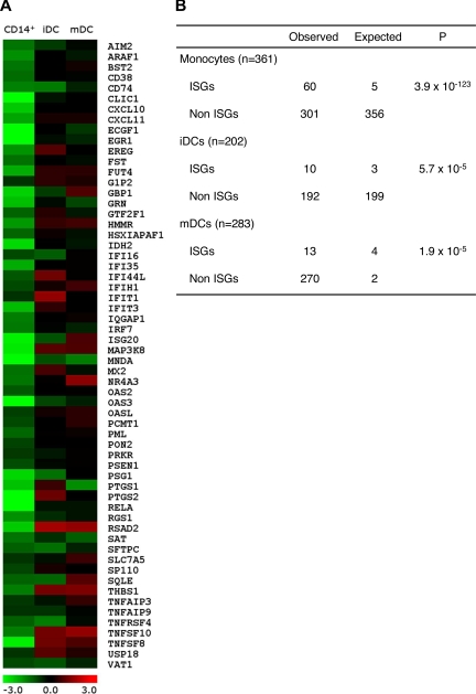
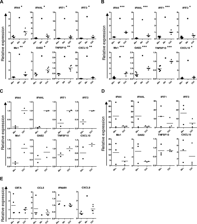
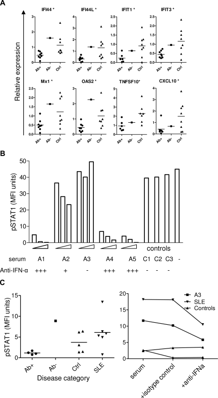
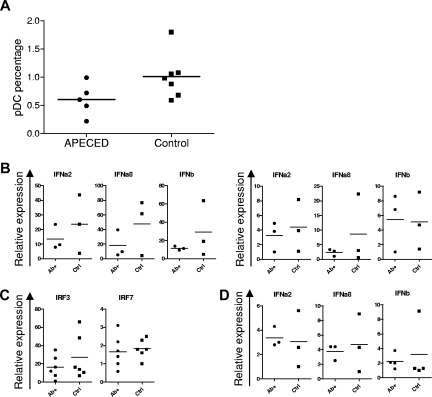
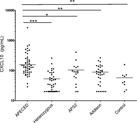
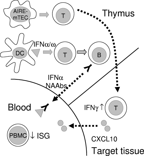
Neutralizing autoantibodies to type I, but not type II, interferons (IFNs) are found at high titers in almost every patient with autoimmune polyendocrinopathy candidiasis ectodermal dystrophy (APECED), a disease caused by AIRE gene mutations that lead to defects in thymic T-cell selection. Combining genome-wide expression array with real time RT-PCR assays, we here demonstrate that antibodies against IFN-alpha cause highly significant down-regulation of interferon-stimulated gene expression in cells from APECED patients' blood by blocking their highly dilute endogenous IFNs. This down-regulation was lost progressively as these APECED cells matured in cultures without neutralizing autoantibodies. Most interestingly, a rare APECED patient with autoantibodies to IFN-omega but not IFN-alpha showed a marked increase in expression of the same interferon-stimulated genes. We also report unexpected increases in serum CXCL10 levels in APECED. Our results argue that the breakdown of tolerance to IFNs in AIRE deficiency is associated with impaired responses to them in thymus, and highlight APECED as another autoimmune disease with associated dysregulation of IFN activity.


Figures

Figure 1
Figure 1
Microarray analysis of APECED and control monocytes and monocyte-derived DCs. (A) The gene expression profiles (log2 of fold change between APECED patient A8 and control 2) of 61 differentially expressed ISGs in monocytes, iDCs, and mDCs. Most of the ISGs lost their differential expression by the iDC stage. (B) The differential expression of ISGs in APECED patient monocytes, iDCs, and mDCs. n indicates the total number of genes down-regulated 1.5-fold in each microarray data set. ISGs and non-ISGs indicate the number of genes, respectively, characterized or not characterized as ISGs. The expected numbers of ISGs and non-ISGs were calculated assuming the random selection. χ2 test was used to calculate the P values to determine whether the deviation is significant.
Figure 2
Figure 2
Decreased expression of ISGs in APECED cell populations. Expression of ISGs (A) and other genes (E) in freshly isolated PBMCs from IFN-α antibody-positive (Ab+, 4 cases) and -negative (Ab, 1 case) APECED patients and healthy controls (Ctrl, 5 cases). (C) Expression of ISGs in freshly isolated plasmacytoid DCs (2 cases in each group). (B) Expression of ISGs in freshly isolated monocytes from IFN-α antibody-positive (6 cases) and -negative (1 case) APECED patients and healthy controls (7 cases). (D) Expression of ISGs in monocyte-derived DCs from APECED patients and healthy controls (3 cases in each group). *P < .05, **P < .001, ***P < .001, comparison of the Ab+ APECED patient and control blood cells. Bars represent group averages.
Figure 3
Figure 3
Effect of APECED sera on ISG expression and STAT1 phosphorylation. (A) Expression of ISGs in control monocytes incubated in 20% autologous sera with the addition of 2% APECED sera-positive (Ab+) or -negative (Ab) for IFN-α antibodies, or with control serum (Ctrl) for 18 hours. *P < .05 between IFN-α antibody–positive APECED patients and healthy controls. (B) U937 cells were treated with 1000 U/mL IFN-α for 15 minutes or with the same concentration of IFN-α preincubated with 2%, 5%, or 10% of APECED (A1-A5) or 10% of control (C1-C3) sera, stained for intacellular pSTAT1 and measured by flow cytometry. (C) Control PBMCs were incubated with 50% of serum samples from APECED patients positive (Ab+) or negative (Ab) for IFN-α antibodies, healthy controls (Ctrl), or SLE patients for 15 minutes and stained for intracellular pSTAT1 to test for IFN activity in the sera (left panel). Control PBMCs were incubated with 50% of serum samples from an APECED patient negative for IFN-α autoantibodies (A3), an SLE patient with IFN activity, or two healthy controls for 15 minutes with or without the addition of neutralizing anti–IFN-α antibody or isotype control antibody as indicated, and stained for intracellular pSTAT1 (right panel).
Figure 4
Figure 4
Normal plasmacytoid DC numbers and dendritic cell IFN production in APECED. (A) Percentages of plasmacytoid DCs (pDCs) among PBMCs in APECED and control groups (P = .079, t test). (B) Expression of type I IFN genes after plasmacytoid DC stimulation for 6 hours with live influenza virus (left panel) or 2.5 μM CpG (right panel) in IFN-α–positive APECED (Ab+) or control persons (Ctrl). (C) IRF3 and IRF7 expression in stimulated plasmacytoid DCs from APECED or control individuals. (D) Type I IFN gene expression was studied after 18 hours of stimulation of monocyte-derived DCs from APECED patients (Ab+) and healthy controls (Ctrl) with 25 μg/mL of poly(I:C).
Figure 5
Figure 5
CXCL10 levels in APECED and control sera. CXCL10 levels are significantly higher in APECED than in unaffected AIRE-heterozygous relatives, patients with APS2, Addison disease, or healthy controls. Geometric means of groups are indicated on plots. *P < .05, **P < .01, ***P < .001.
Figure 6
Figure 6
Schematic illustration of thymic and peripheral events in APECED. Tissue-restricted antigen-specific T cells are not deleted in APECED thymus because of the deficient function of AIRE in medullary thymic epithelial cells (mTEC), and escape to cause tissue damage in endocrine organs in APECED patients. We speculate that, because of aberrant cell death or alternative danger signal, human AIRE-deficient thymus (and thymomas) assume some functions of secondary lymphoid organs, with associated overproduction of type I IFNs by thymic DCs. These DCs are activated and present a broad spectrum of type I interferons to autoimmunize specific T and then B cells. The resulting high titer neutralizing antibodies inhibit ISG responses of PBMCs to basal circulating levels of type I IFNs. Autoreactive T cells infiltrating target endocrine tissues secrete IFN-γ. This induces the production of CXCL10 and further expansion of the adjacent infiltrates.

References

    1. Pestka S, Krause CD, Walter MR. Interferons, interferon-like cytokines, and their receptors. Immunol Rev. 2004;202:8–32. - PubMed
    1. Baccala R, Kono DH, Theofilopoulos AN. Interferons as pathogenic effectors in autoimmunity. Immunol Rev. 2005;204:9–26. - PubMed
    1. Saksela E, Virtanen I, Hovi T, Secher DS, Cantell K. Monocyte is the main producer of human leukocyte alpha interferons following Sendai virus induction. Prog Med Virol. 1984;30:78–86. - PubMed
    1. Liu YJIPC: professional type 1 interferon-producing cells and plasmacytoid dendritic cell precursors. Annu Rev Immunol. 2005;23:275–306. - PubMed
    1. Blanco P, Palucka AK, Pascual V, Banchereau J. Dendritic cells and cytokines in human inflammatory and autoimmune diseases. Cytokine Growth Factor Rev. 2008;19:41–52. - PMC - PubMed

Publication types

MeSH terms