

Impaired purinergic neurotransmission to mesenteric arteries in deoxycorticosterone acetate-salt hypertensive rats
- PMID: 18606906
- PMCID: PMC2748660
- DOI: 10.1161/HYPERTENSIONAHA.108.110353
Impaired purinergic neurotransmission to mesenteric arteries in deoxycorticosterone acetate-salt hypertensive rats
Abstract
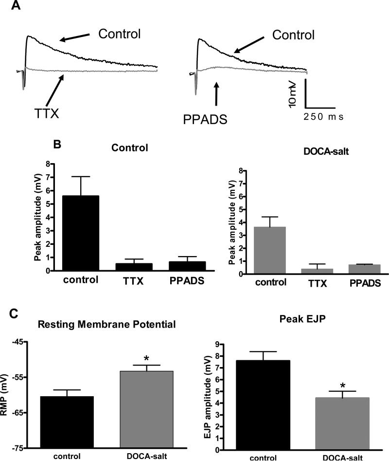
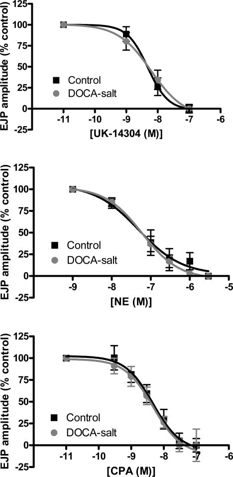
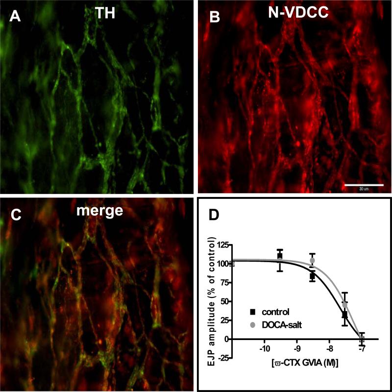
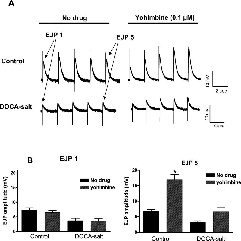
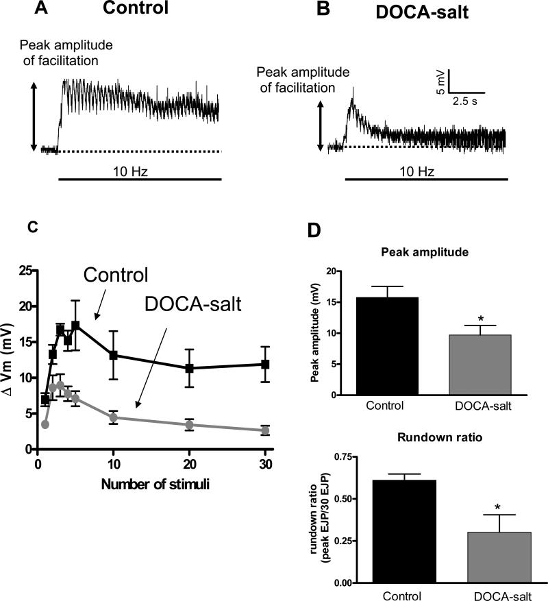
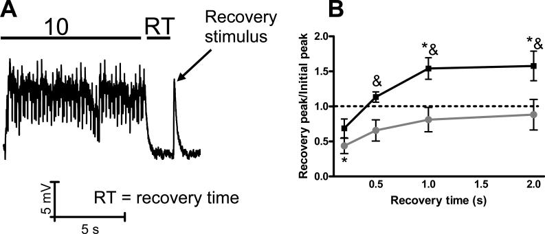
Sympathetic nerves release norepinephrine and ATP onto mesenteric arteries. In deoxycorticosterone acetate (DOCA)-salt hypertensive rats, there is increased arterial sympathetic neurotransmission attributable, in part, to impaired prejunctional regulation of norepinephrine release. Prejunctional regulation purinergic transmission in hypertension is less well understood. We hypothesized that alpha(2)-adrenergic receptor dysfunction alters purinergic neurotransmission to arteries in DOCA-salt hypertensive rats. Mesenteric artery preparations were maintained in vitro, and intracellular electrophysiological methods were used to record excitatory junction potentials (EJPs) from smooth muscle cells. EJP amplitude was reduced in smooth muscle cells from DOCA-salt (4+/-1 mV) compared with control arteries (9+/-1 mV; P<0.05). When using short trains of stimulation (0.5 Hz; 5 pulses), the alpha(2)adrenergic receptor antagonist yohimbine (1 micromol/L) potentiated EJPs in control more than in DOCA-salt arteries (180+/-35% versus 86+/-7%; P<0.05). Norepinephrine (0.1 to 3.0 micromol/L), the alpha(2)adrenergic receptor agonist UK 14304 (0.001 to 0.100 micromol/L), the A(1) adenosine receptor agonist cyclopentyladensosine (0.3 to 100.0 micromol/L), and the N-type calcium channel blocker omega-conotoxin GVIA (0.0003 to 0.1000 micromol/L) decreased EJP amplitude equally well in control and DOCA-salt arteries. Trains of stimuli (10 Hz) depleted ATP stores more completely, and the latency to EJP recovery was longer in DOCA-salt compared with control arteries. These data indicate that there is reduced purinergic input to mesenteric arteries of DOCA-salt rats because of decreased ATP bioavailability in sympathetic nerves. These data highlight the potential importance of impaired purinergic regulation of arterial tone as a target for drug treatment of hypertension.
Figures
Similar articles
-
Antioxidant treatment restores prejunctional regulation of purinergic transmission in mesenteric arteries of deoxycorticosterone acetate-salt hypertensive rats.Neuroscience. 2010 Jun 30;168(2):335-45. doi: 10.1016/j.neuroscience.2010.03.061. Epub 2010 Apr 14. Neuroscience. 2010. PMID: 20398741 Free PMC article.
-
Impaired function of prejunctional adenosine A1 receptors expressed by perivascular sympathetic nerves in DOCA-salt hypertensive rats.J Pharmacol Exp Ther. 2013 Apr;345(1):32-40. doi: 10.1124/jpet.112.199612. Epub 2013 Feb 8. J Pharmacol Exp Ther. 2013. PMID: 23397055 Free PMC article.
-
Alterations in sympathetic neuroeffector transmission to mesenteric arteries but not veins in DOCA-salt hypertension.Auton Neurosci. 2010 Jan 15;152(1-2):11-20. doi: 10.1016/j.autneu.2009.08.008. Epub 2009 Nov 13. Auton Neurosci. 2010. PMID: 19914150 Free PMC article.
-
Impaired function of alpha2-adrenergic autoreceptors on sympathetic nerves associated with mesenteric arteries and veins in DOCA-salt hypertension.Am J Physiol Heart Circ Physiol. 2004 Apr;286(4):H1558-64. doi: 10.1152/ajpheart.00592.2003. Epub 2003 Dec 11. Am J Physiol Heart Circ Physiol. 2004. PMID: 14670814
-
A review of deoxycorticosterone acetate-salt hypertension and its relevance for cardiovascular physiotherapy research.J Phys Ther Sci. 2015 Jan;27(1):303-7. doi: 10.1589/jpts.27.303. Epub 2015 Jan 9. J Phys Ther Sci. 2015. PMID: 25642096 Free PMC article. Review.
Cited by
-
Localization of NADPH oxidase in sympathetic and sensory ganglion neurons and perivascular nerve fibers.Auton Neurosci. 2009 Dec 3;151(2):90-7. doi: 10.1016/j.autneu.2009.07.010. Epub 2009 Aug 27. Auton Neurosci. 2009. PMID: 19716351 Free PMC article.
-
Antioxidant treatment restores prejunctional regulation of purinergic transmission in mesenteric arteries of deoxycorticosterone acetate-salt hypertensive rats.Neuroscience. 2010 Jun 30;168(2):335-45. doi: 10.1016/j.neuroscience.2010.03.061. Epub 2010 Apr 14. Neuroscience. 2010. PMID: 20398741 Free PMC article.
-
Impaired function of prejunctional adenosine A1 receptors expressed by perivascular sympathetic nerves in DOCA-salt hypertensive rats.J Pharmacol Exp Ther. 2013 Apr;345(1):32-40. doi: 10.1124/jpet.112.199612. Epub 2013 Feb 8. J Pharmacol Exp Ther. 2013. PMID: 23397055 Free PMC article.
-
Sympathetic overdrive in obesity involves purinergic hyperactivity in the resistance vasculature.J Physiol. 2011 Jul 1;589(Pt 13):3289-307. doi: 10.1113/jphysiol.2011.207944. Epub 2011 May 16. J Physiol. 2011. PMID: 21576274 Free PMC article.
-
Alterations in sympathetic neuroeffector transmission to mesenteric arteries but not veins in DOCA-salt hypertension.Auton Neurosci. 2010 Jan 15;152(1-2):11-20. doi: 10.1016/j.autneu.2009.08.008. Epub 2009 Nov 13. Auton Neurosci. 2010. PMID: 19914150 Free PMC article.
References
-
- King AJ, Osborn JW, Fink GD. Splanchnic circulation is a critical neural target in angiotensin II salt hypertension in rats. Hypertension. 2007;50(3):547–56. - PubMed
-
- Donoso MV, Steiner M, Huidobro-Toro JP. BIBP 3226, suramin and prazosin identify neuropeptide Y, adenosine 5'-triphosphate and noradrenaline as sympathetic cotransmitters in the rat arterial mesenteric bed. J Pharmacol Exp Ther. 1997;282(2):691–8. - PubMed
-
- Bao JX, Gonon F, Stjarne L. Kinetics of ATP- and noradrenaline-mediated sympathetic neuromuscular transmission in rat tail artery. Acta Physiol Scand. 1993;149(4):503–19. - PubMed
-
- Lundberg JM, et al. High levels of neuropeptide Y in peripheral noradrenergic neurons in various mammals including man. Neurosci Lett. 1983;42(2):167–72. - PubMed
Publication types
MeSH terms
Substances
Grants and funding
LinkOut - more resources
Full Text Sources
Medical
