

Impact of white matter hyperintensity volume progression on rate of cognitive and motor decline
- PMID: 18606964
- PMCID: PMC2676966
- DOI: 10.1212/01.wnl.0000316799.86917.37
Impact of white matter hyperintensity volume progression on rate of cognitive and motor decline
Abstract
Background: White matter hyperintensity (WMH) change on brain MRI is observed with increased frequency in the elderly and has been independently associated with neurologic decline. The degree to which the location and rate of volume increase in WMH affects other structural brain changes along with cognitive and motor performance over time may determine subsequent degrees of risk for dementia and other syndromes of aging.
Methods: One hundred four cognitively intact men and women followed longitudinally for up to 13 years underwent at least three MRIs with corresponding annual cognitive and neurologic assessments. Brain volume, ventricular CSF (vCSF), and total periventricular (PV) and subcortical WMH volumes were measured. Progression of MRI volumes was examined in relation to rates of cognitive, motor, and cerebral volume change based on slopes of outcomes.
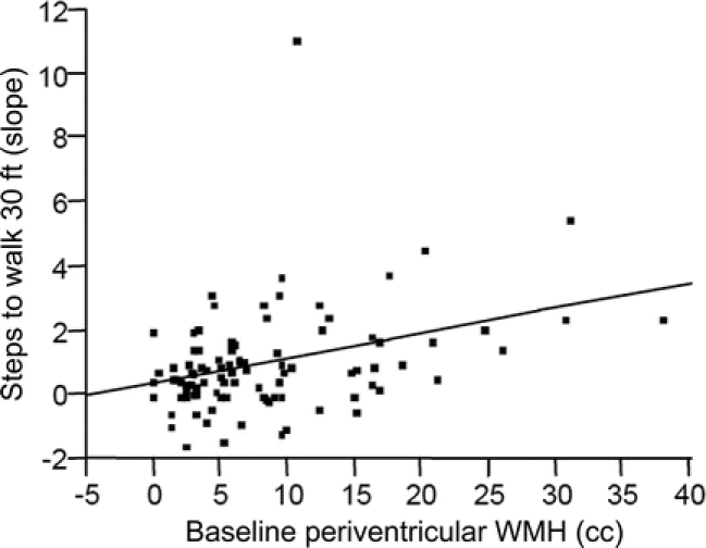
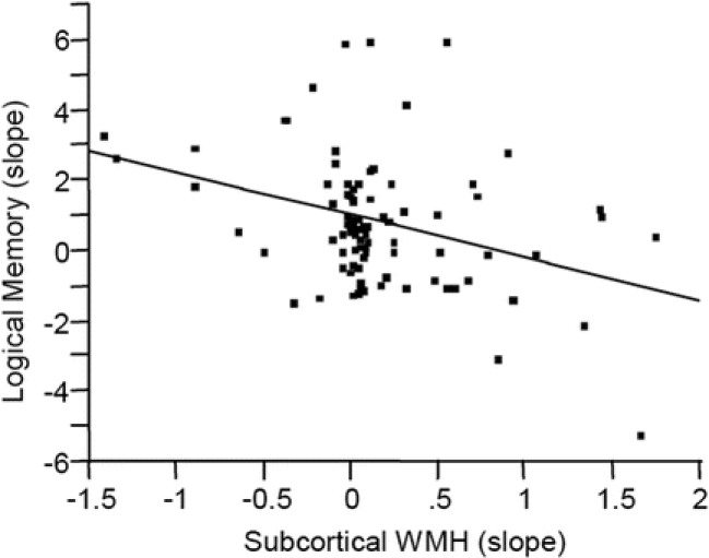
Results: Higher initial total and PV WMH volume was associated with total WMH, PV WMH, and vCSF progression, and with increased time and number of steps to walk 30 feet. Progression of PV WMH volume was associated with increased time to walk 30 feet. Progression of subcortical WMH volume was associated with decreased performance on logical memory testing and increased rate of vCSF volume change.
Conclusion: Increased total and periventricular (PV) white matter hyperintensity (WMH) burden and progression of PV WMH burden are associated with decreased gait performance over time, while progression of subcortical WMH volume is associated with memory decline in cognitively intact elderly. Greater progression of WMH burden is associated with an increased risk of memory and gait dysfunction, and thus should not be considered a benign process.
Figures
References
-
- Yue NC, Arnold AM, Longstreth WT Jr, et al. Sulcal, ventricular, and white matter changes at MR imaging in the aging brain: data from the cardiovascular health study [see comment]. Radiology 1997;202:33–39. - PubMed
-
- Baum KA, Schulte C, Girke W, Reischies FM, Felix R. Incidental white-matter foci on MRI in “healthy” subjects: evidence of subtle cognitive dysfunction. Neuroradiol 1996;38:755–760. - PubMed
-
- DeCarli C, Murphy DG, Tranh M, et al. The effect of white matter hyperintensity volume on brain structure, cognitive performance, and cerebral metabolism of glucose in 51 healthy adults. Neurology 1995;45:2077–2084. - PubMed
-
- Junque C, Pujol J, Vendrell P, et al. Leuko-araiosis on magnetic resonance imaging and speed of mental processing. Arch Neurol 1990;47:151–156. - PubMed
-
- Longstreth WT Jr, Manolio TA, Arnold A, et al. Clinical correlates of white matter findings on cranial magnetic resonance imaging of 3301 elderly people: The Cardiovascular Health Study. [see comment]. Stroke 1996;27:1274–1282. - PubMed
MeSH terms
LinkOut - more resources
Full Text Sources
Medical