

Crystallization and preliminary X-ray characterization of the genetically encoded fluorescent calcium indicator protein GCaMP2
- PMID: 18607093
- PMCID: PMC2443961
- DOI: 10.1107/S1744309108016059
Crystallization and preliminary X-ray characterization of the genetically encoded fluorescent calcium indicator protein GCaMP2
Abstract
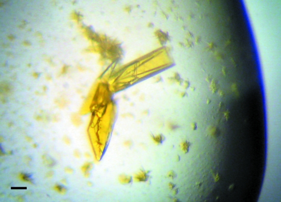
Fluorescent proteins and their engineered variants have played an important role in the study of biology. The genetically encoded calcium-indicator protein GCaMP2 comprises a circularly permuted fluorescent protein coupled to the calcium-binding protein calmodulin and a calmodulin target peptide, M13, derived from the intracellular calmodulin target myosin light-chain kinase and has been used to image calcium transients in vivo. To aid rational efforts to engineer improved variants of GCaMP2, this protein was crystallized in the calcium-saturated form. X-ray diffraction data were collected to 2.0 A resolution. The crystals belong to space group C2, with unit-cell parameters a = 126.1, b = 47.1, c = 68.8 A, beta = 100.5 degrees and one GCaMP2 molecule in the asymmetric unit. The structure was phased by molecular replacement and refinement is currently under way.
Figures


References
-
- Chalfie, M., Tu, Y., Euskirchen, G., Ward, W. W. & Prasher, D. C. (1994). Science, 263, 802–805. - PubMed
-
- Collaborative Computational Project, Number 4 (1994). Acta Cryst. D50, 760–763. - PubMed
-
- Diez-Garcia, J., Matsushita, S., Mutoh, H., Nakai, J., Ohkura, M., Yokoyama, J., Dimitrov, D. & Knopfel, T. (2005). Eur. J. Neurosci.22, 627–635. - PubMed
-
- Giepmans, B. N., Adams, S. R., Ellisman, M. H. & Tsien, R. Y. (2006). Science, 312, 217–224. - PubMed
Publication types
MeSH terms
Substances
Grants and funding
LinkOut - more resources
Full Text Sources
Other Literature Sources
Miscellaneous
