

The rare association of moyamoya disease and cerebral arteriovenous malformations: a case report
- PMID: 18607130
- PMCID: PMC2627196
- DOI: 10.3348/kjr.2008.9.s.s65
The rare association of moyamoya disease and cerebral arteriovenous malformations: a case report
Abstract
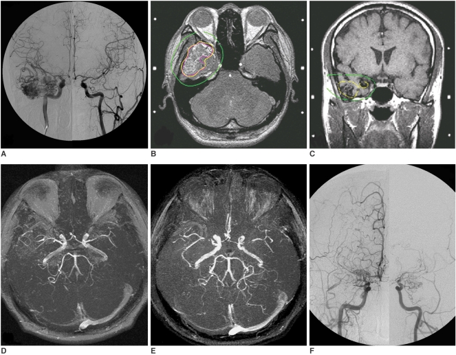
A 36-year-old man was diagnosed with a right temporal lobe grade II cerebral arteriovenous malformation (cAVM) and was treated with radiosurgery. At nine months after the cAVM radiosurgery, the patient began to develop bilateral focal narrowing at the M1 segments of the bilateral middle cerebral arteries. The narrowing progressively deteriorated as was demonstrated on longitudinal serial follow-up MR imaging. X-ray angiography performed at 51 months after radiosurgery confirmed that the cAVM was cured and a diagnosis of moyamoya disease. To the best of our knowledge, this is the first case of cAVM-associated moyamoya disease that developed after radiosurgery. Given the chronological sequence of disease development and radiation dose distribution of radiosurgery, it is proposed that humoral or unknown predisposing factors, rather than direct radiation effects, are the cause of moyamoya disease associated with cAVM.
Figures
Similar articles
-
Embolization of arteriovenous fistula after radiosurgery for multiple cerebral arteriovenous malformations.Kaohsiung J Med Sci. 2005 Dec;21(12):571-7. doi: 10.1016/S1607-551X(09)70210-4. Kaohsiung J Med Sci. 2005. PMID: 16670050 Free PMC article.
-
Radiosurgical treatment of a cerebral arteriovenous malformation in a patient with moyamoya disease: case report.Neurosurgery. 2002 Aug;51(2):478-81; discussion 481-2. Neurosurgery. 2002. PMID: 12182787 Review.
-
Leksell Gamma Knife for pediatric and adolescent cerebral arteriovenous malformations: results of 100 cases followed up for at least 36 months.J Neurosurg Pediatr. 2015 Dec;16(6):736-47. doi: 10.3171/2015.4.PEDS158. Epub 2015 Sep 4. J Neurosurg Pediatr. 2015. PMID: 26339954
-
Moyamoya syndrome associated with γ knife surgery for cerebral arteriovenous malformation: case report.Neurol Med Chir (Tokyo). 2012;52(5):343-5. doi: 10.2176/nmc.52.343. Neurol Med Chir (Tokyo). 2012. PMID: 22688073
-
Chronic encapsulated intracerebral hematoma formation after radiosurgery for cerebral arteriovenous malformation.Neurol India. 2011 Jul-Aug;59(4):624-6. doi: 10.4103/0028-3886.84352. Neurol India. 2011. PMID: 21891948 Review.
Cited by
-
Moyamoya disease and arteriovenous fistula of the epiaortic vessels.Neurol Sci. 2010 Dec;31(6):821-4. doi: 10.1007/s10072-010-0331-4. Epub 2010 Jun 5. Neurol Sci. 2010. PMID: 20526643
-
An Uncommon Case of Moyamoya Syndrome Is Accompanied by an Arteriovenous Malformation with the Involvement of Dural Arteries.Int J Mol Sci. 2023 Mar 21;24(6):5911. doi: 10.3390/ijms24065911. Int J Mol Sci. 2023. PMID: 36982983 Free PMC article.
-
Moyamoya disease associated with arteriovenous malformation and anterior communicating artery aneurysm: A case report and literature review.Exp Ther Med. 2016 Jul;12(1):267-271. doi: 10.3892/etm.2016.3289. Epub 2016 Apr 21. Exp Ther Med. 2016. PMID: 27347048 Free PMC article.
References
-
- Mawad ME, Hilal SK, Michelsen WJ, Stein B, Ganti SR. Occlusive vascular disease associated with cerebral arteriovenous malformations. Radiology. 1984;153:401–408. - PubMed
-
- Enam SA, Malik GM. Association of cerebral arteriovenous malformations and spontaneous occlusion of major feeding arteries: clinical and therapeutic implications. Neurosurgery. 1999;45:1105–1111. - PubMed
-
- Nakashima T, Nakayama N, Furuichi M, Kokuzawa J, Murukawa T, Sakai N. Arteriovenous malformation in association with moyamoya disease Report of two cases. Neurosurg Focus. 1998;5:E6. - PubMed
-
- Halatsch ME, Rustenbeck HH, Jansen J. Progression of arteriovenous malformation in moyamoya syndrome. Acta Neurochir (Wien) 1997;139:82–85. - PubMed
Publication types
MeSH terms
LinkOut - more resources
Full Text Sources
