Review
. 2008 Sep;466(9):2168-75.
doi: 10.1007/s11999-008-0338-9. Epub 2008 Jul 8.

Specific tyrosine kinase inhibitors regulate human osteosarcoma cells in vitro

Affiliations
Review

Specific tyrosine kinase inhibitors regulate human osteosarcoma cells in vitro

Patrick J Messerschmitt et al. Clin Orthop Relat Res. 2008 Sep.

Abstract

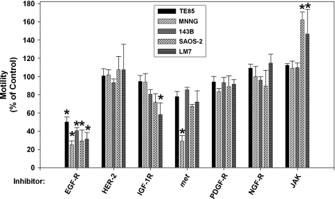
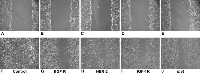
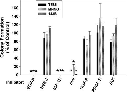
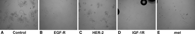
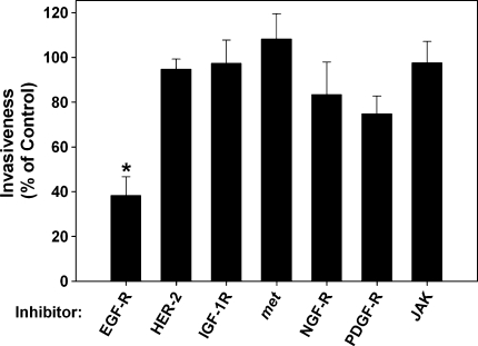
Inhibitors of specific tyrosine kinases are attractive lead compounds for development of targeted chemotherapies for many tumors, including osteosarcoma. We asked whether inhibition of specific tyrosine kinases would decrease the motility, colony formation, and/or invasiveness by human osteosarcoma cell lines (TE85, MNNG, 143B, SAOS-2, LM-7). An EGF-R inhibitor reduced motility of all five cell lines by 50% to 80%. In contrast, an IGF-1R inhibitor preferentially reduced motility by 42% in LM-7 cells and a met inhibitor preferentially reduced motility by 80% in MNNG cells. The inhibitors of EGF-R, IGF-1R, and met reduced colony formation by more than 80% in all tested cell lines (TE85, MNNG, 143B). The EGF-R inhibitor reduced invasiveness by 62% in 143B cells. The JAK inhibitor increased motility of SAOS-2 and LM7 cells without affecting colony formation or invasiveness. Inhibitors of HER-2, NGF-R, and PDGF-Rs did not affect motility, invasiveness, or colony formation. These results support the hypothesis that specific tyrosine kinases regulate tumorigenesis and/or metastasis in osteosarcoma.


Figures

Fig. 1
Fig. 1
Specific tyrosine kinase inhibitors reduced motility by the osteosarcoma cell lines. Motility was measured 4 hours after scraping in the presence of the tyrosine kinase inhibitors listed in Table 1 or 1% DMSO as a vehicle control. Bars represent the means ± standard error of mean of four to six independent experiments, each with four to six scrapes per group. Asterisks denote differences in motility compared with the control group without inhibitor (EGF-R inhibitor: TE85 [p = 9.4 × 10−9], MNNG [p = 1.0 × 10−10], 143B [p = 1.0 × 10−10], SAOS-2 [p = 6.0 × 10−9], LM7 [p = 1.0 × 10−10]’ IGF-1R inhibitor: LM7 [p = 1.6 × 10−4]; met inhibitor: MNNG [p = 1.0 × 10−10]; JAK inhibitor: SAOS-2 [p = 3.6 × 10−8], LM7 [p = 2.8 × 10−5]).
Fig. 2A–J
Fig. 2A–J
Photomicrographs show the effects of specific tyrosine kinase inhibitors on motility. Images of the same microscopic field of the 143B cell line at the time of initial scraping (A–E) and after culture for 4 hours in the presence of 1% DMSO as a vehicle control (F), an inhibitor of EGF-R (G), an inhibitor of HER-2 (H), an inhibitor of IGF-1R (I), and an inhibitor of met (J).
Fig. 3
Fig. 3
Specific tyrosine kinase inhibitors reduced colony formation by the osteosarcoma cell lines. Colony formation assays were performed in the presence of the tyrosine kinase inhibitors listed in Table 1 or 1% DMSO as a vehicle control. Bars represent the means ± standard error of mean of three individual experiments, each with three wells per group. Asterisks denote a decrease in the colony count compared with the control group without inhibitor (p = 9.2 × 10−9 for each asterisk except: 143B cells in the presence of the met inhibitor [p = 1.04 × 10−8] and MNNG cells in the presence of the met inhibitor [p = 9.9 × 10−7]).
Fig. 4A–E
Fig. 4A–E
Photomicrographs show the effects of specific tyrosine kinase inhibitors on colony formation. Images of the 143B cell lines were collected after culture for 4 days in the presence of 1% DMSO as a vehicle control (A), an inhibitor of EGF-R (B), an inhibitor of HER-2 (C), an inhibitor of IGF-1R (D), and an inhibitor of met (E). (D–E) reveal air bubbles, which became trapped in the collagen gel.
Fig. 5
Fig. 5
Specific tyrosine kinase inhibitors reduced invasiveness by the 143B cell line. Basement membrane invasion assays were performed in the presence of the tyrosine kinase inhibitors listed in Table 1 or 1% DMSO as a vehicle control. Bars represent the means ± standard error of mean of three to five individual experiments, each with five wells per group. The asterisk denotes a decrease (p = 6.1 × 10−5) in invasiveness compared with the control group without inhibitor.

References

    1. {'text': '', 'ref_index': 1, 'ids': [{'type': 'DOI', 'value': '10.1159/000058533', 'is_inner': False, 'url': 'https://doi.org/10.1159/000058533'}, {'type': 'PubMed', 'value': '11408805', 'is_inner': True, 'url': 'https://pubmed.ncbi.nlm.nih.gov/11408805/'}]}
    2. Akatsuka T, Wada T, Kokai Y, Sawada N, Yamawaki S, Ishii S. Loss of ErbB2 expression in pulmonary metastatic lesions in osteosarcoma. Oncology. 2001;60:361–366. - PubMed
    1. {'text': '', 'ref_index': 1, 'ids': [{'type': 'PubMed', 'value': '2438036', 'is_inner': True, 'url': 'https://pubmed.ncbi.nlm.nih.gov/2438036/'}]}
    2. Albini A, Iwamoto Y, Kleinman HK, Martin GR, Aaronson SA, Kozlowski JM, McEwan RN. A rapid in vitro assay for quantitating the invasive potential of tumor cells. Cancer Res. 1987;47:3239–3245. - PubMed
    1. {'text': '', 'ref_index': 1, 'ids': [{'type': 'DOI', 'value': '10.1042/BJ20070797', 'is_inner': False, 'url': 'https://doi.org/10.1042/bj20070797'}, {'type': 'PMC', 'value': 'PMC2267365', 'is_inner': False, 'url': 'https://pmc.ncbi.nlm.nih.gov/articles/PMC2267365/'}, {'type': 'PubMed', 'value': '17850214', 'is_inner': True, 'url': 'https://pubmed.ncbi.nlm.nih.gov/17850214/'}]}
    2. Bain J, Plater L, Elliott M, Shpiro N, Hastie CJ, McLauchlan H, Klevernic I, Arthur JS, Alessi DR, Cohen P. The selectivity of protein kinase inhibitors: a further update. Biochem J. 2007;408:297–315. - PMC - PubMed
    1. {'text': '', 'ref_index': 1, 'ids': [{'type': 'DOI', 'value': '10.1126/science.1125951', 'is_inner': False, 'url': 'https://doi.org/10.1126/science.1125951'}, {'type': 'PubMed', 'value': '16728632', 'is_inner': True, 'url': 'https://pubmed.ncbi.nlm.nih.gov/16728632/'}]}
    2. Baselga J. Targeting tyrosine kinases in cancer: the second wave. Science. 2006;312:1175–1178. - PubMed
    1. {'text': '', 'ref_index': 1, 'ids': [{'type': 'DOI', 'value': '10.1002/(SICI)1097-0215(19990209)80:4<581::AID-IJC16>3.0.CO;2-O', 'is_inner': False, 'url': 'https://doi.org/10.1002/(sici)1097-0215(19990209)80:4<581::aid-ijc16>3.0.co;2-o'}, {'type': 'PubMed', 'value': '9935160', 'is_inner': True, 'url': 'https://pubmed.ncbi.nlm.nih.gov/9935160/'}]}
    2. Benini S, Baldini N, Manara M, Chano T, Serra M, Rizzi S, Lollini P, Picci P, Scotlandi K. Redundancy of autocrine loops in human osteosarcoma cells. Int J Cancer. 1999;80:581–588. - PubMed

Publication types

MeSH terms

Substances