. 2008 Dec;23(6):931-45.
doi: 10.1080/14756360701810082.

A high density assay format for the detection of novel cytotoxic agents in large chemical libraries

Affiliations

A high density assay format for the detection of novel cytotoxic agents in large chemical libraries

David Shum et al. J Enzyme Inhib Med Chem. 2008 Dec.

Abstract

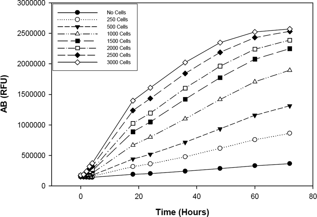
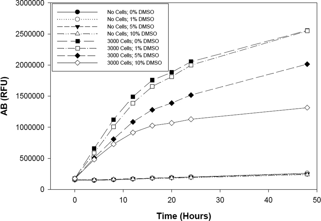
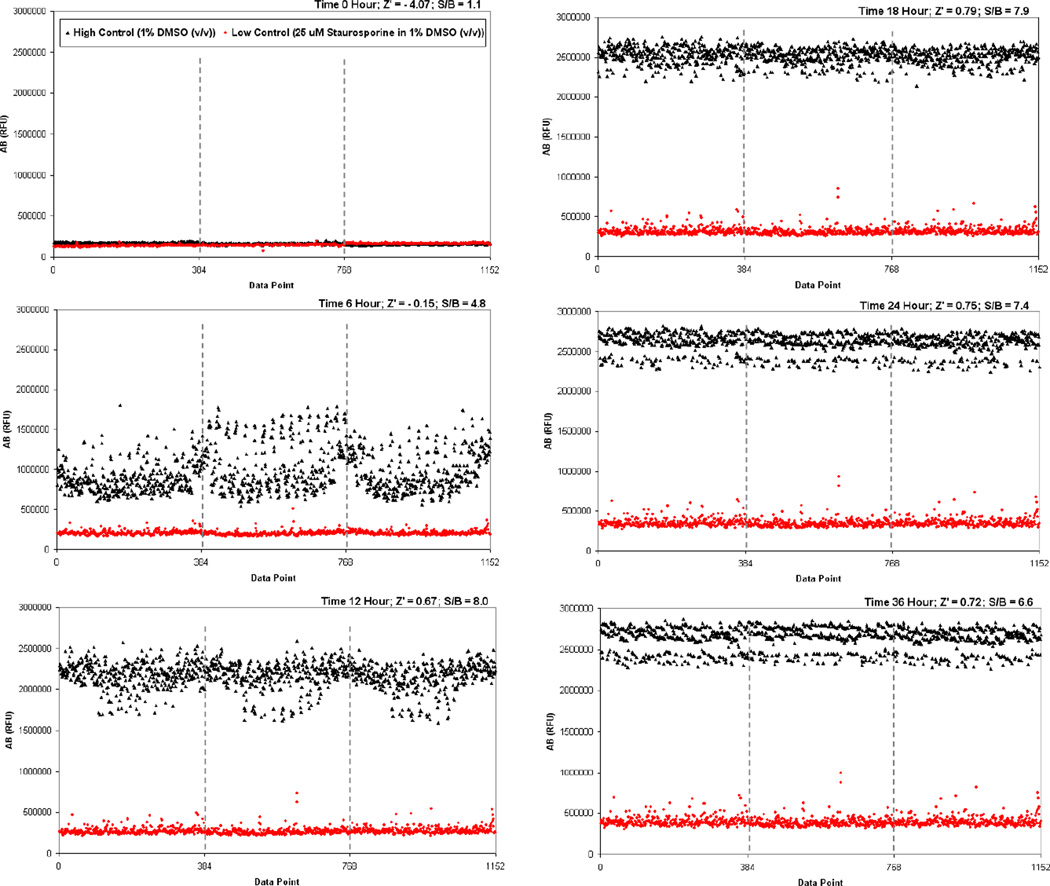
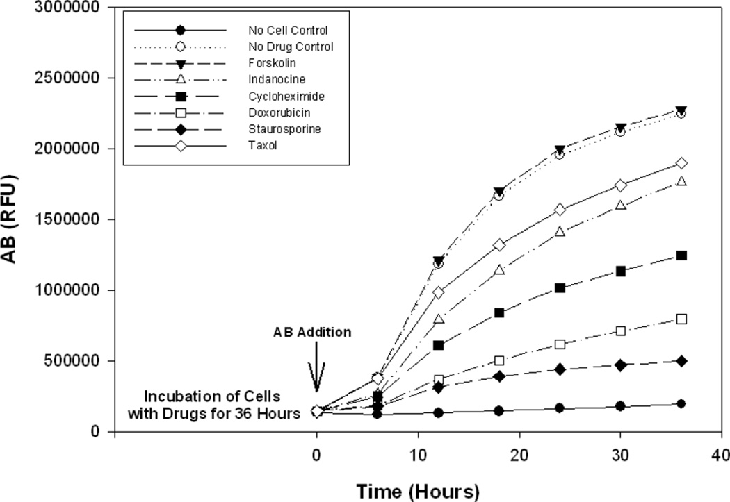
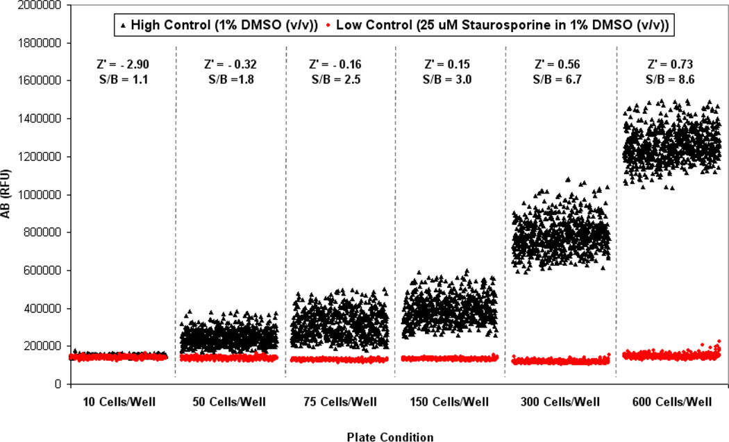
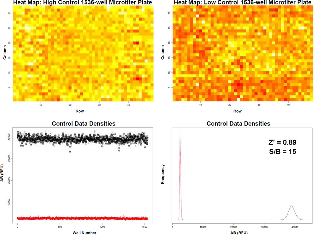
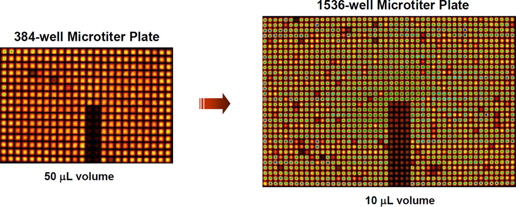
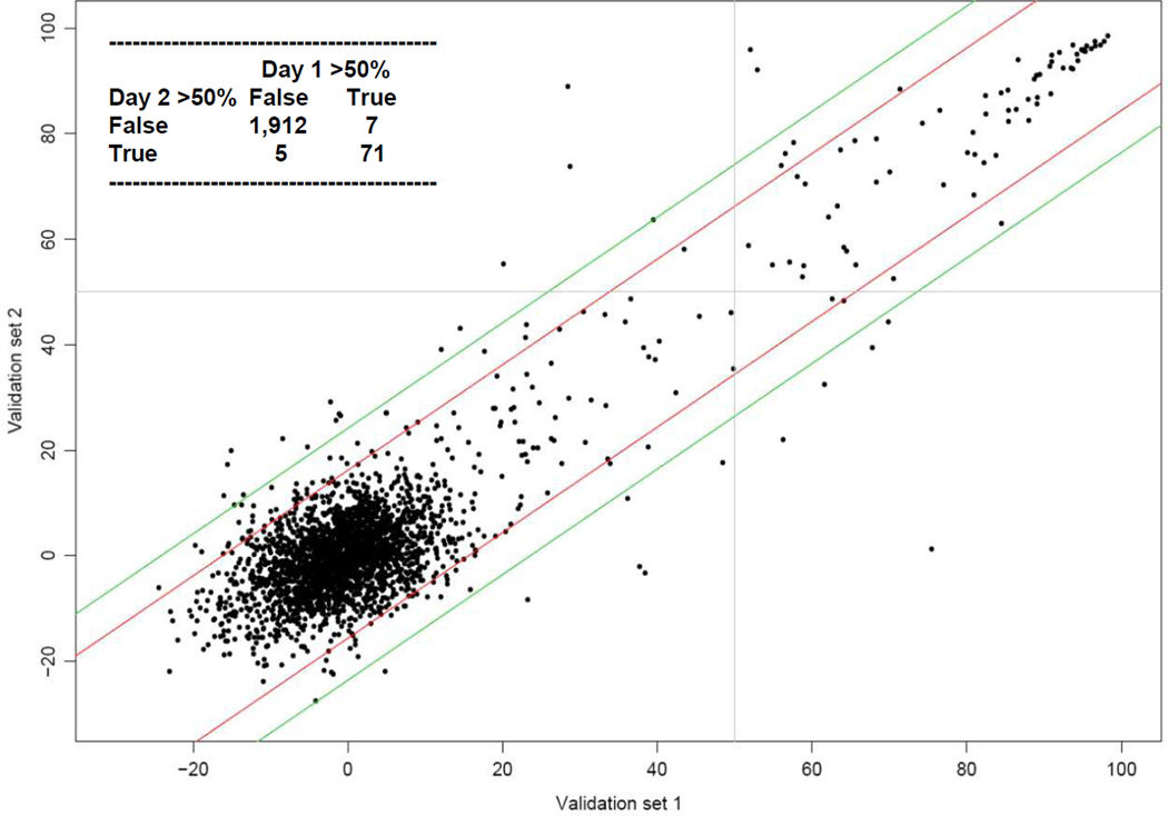
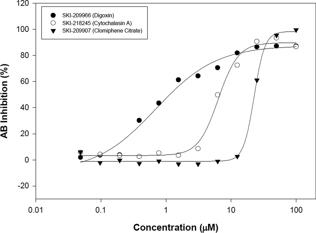
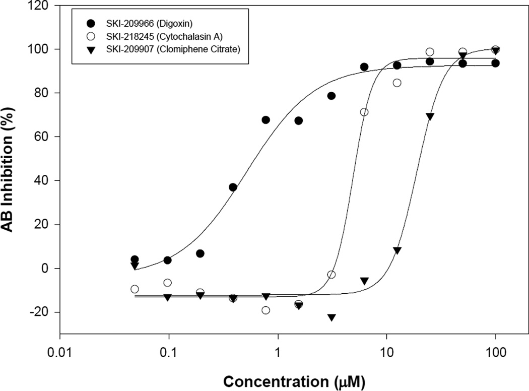
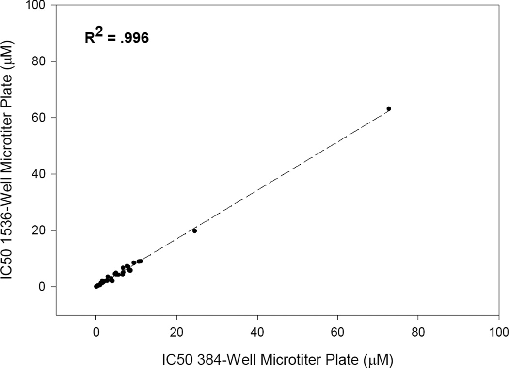
In response to the need for inexpensive high throughput assays for anti-cancer drug screening, a 1536-well microtiter plate based assay utilizing the Alamar Blue fluorescent dye as a measure of cellular growth was validated in 10 microL assay volume. Its robustness was assessed in a screen against a library of 2000 known bioactives; with an overall Z' value of 0.89 for assay robustness, several known cytotoxic agents were identified including and not limited to anthracyclines, cardiac glycosides, gamboges, and quinones. To further test the sensitivity of the assay, IC50 determinations were performed in both 384-well and 1536-well formats and the obtained results show a very good correlation between the two density formats. These findings demonstrate that this newly developed assay is simple to set up, robust, highly sensitive and inexpensive. It could potentially provide a rapid way to screen established and primary tumor cell lines against large chemical libraries.


Figures

Figure 1
Figure 1
a) Growth curve of NCEB1 cell line with various cell densities as assessed by the AB reagent over a time course of 72 hours after AB addition. Cells were seeded into 384-well microtiter plates. An average of 48 wells was used for each data point. b) Effect of DMSO on the growth of NCEB1 cell line as assessed by the AB reagent over a time course of 48 hours. Cells were seeded into 384-well microtiter plates. An average of 96 wells was used for each data point. c) Assessment of assay performance and robustness. Cells were seeded into 384-well microtiter plates and incubated for 36 hours prior to addition of the AB reagent. High control wells contained 1% DMSO (v/v). Low control wells contained 25 µM staurosporine in 1% DMSO (v/v).
Figure 1
Figure 1
a) Growth curve of NCEB1 cell line with various cell densities as assessed by the AB reagent over a time course of 72 hours after AB addition. Cells were seeded into 384-well microtiter plates. An average of 48 wells was used for each data point. b) Effect of DMSO on the growth of NCEB1 cell line as assessed by the AB reagent over a time course of 48 hours. Cells were seeded into 384-well microtiter plates. An average of 96 wells was used for each data point. c) Assessment of assay performance and robustness. Cells were seeded into 384-well microtiter plates and incubated for 36 hours prior to addition of the AB reagent. High control wells contained 1% DMSO (v/v). Low control wells contained 25 µM staurosporine in 1% DMSO (v/v).
Figure 2
Figure 2
Effects of known cytotoxic agents on the growth of the NCEB1 cell line as assessed by the AB reagent over a time course of 36 hours. Cells were seeded into 384-well microtiter plates and incubated for 36 hours prior to the addition of the AB reagent. Negative control wells contained no cells. Positive control wells contained 1% DMSO (v/v). All drugs were screened at a 10 µM final concentration in 1% DMSO (v/v). An average of 48 wells was used for each data point.
Figure 3
Figure 3
Assay miniaturization in 1536-well microtiter plate. Cells were seeded into 1536-well microtiter plates and incubated for 36 hours prior to the addition of the AB reagent and further incubated for 24 hours prior to reading the AB fluorescence. High control wells contained 1% DMSO (v/v). Low control wells contained 25 µM staurosporine in 1% DMSO (v/v).
Figure 4
Figure 4
Control experiment to evaluate assay robustness and performance in 1536-well microtiter format. Top panel shows heat map analysis of the high and low control plate. Bottom panel shows AB signal and distribution of high and low control points. High control wells contained 1% DMSO (v/v). Low control wells contained 25 µM staurosporine in 1% DMSO (v/v).
Figure 5
Figure 5
Sample images of 384-well microtiter plate and 1536-well microtiter plate as imaged by the LEADseeker™ Multimodality Imaging System.
Figure 6
Figure 6
Scatter plot of 2,000 compound validation of the NCEB1 cell line. Scatter plot shows percent inhibition of duplicate values for each compound.
Figure 7
Figure 7
a) IC50 curves of each compound in 384-well microplate format: Digoxin, 0.8 µM; Cytochalasin A, 8.5 µM; and Clomiphene Citrate, 24.5 µM. b) IC50 curves of each compound in 1536-well microplate format: Digoxin, 0.6 µM; Cytochalasin A, 5.8 µM; and Clomiphene Citrate, 19.8 µM. c) Correlation plot of IC50 values in 384-well microtiter plate versus 1536-well microtiter plate. NCEB1 cells were incubated with compounds for 48 hours and then incubated with AB reagent for another 24 hours. High control wells contained 1% DMSO (v/v). Low control wells contained 25 µM staurosporine in 1% DMSO (v/v). IC50 values are an average of two independently fitted dose response curves for each data set.
Figure 7
Figure 7
a) IC50 curves of each compound in 384-well microplate format: Digoxin, 0.8 µM; Cytochalasin A, 8.5 µM; and Clomiphene Citrate, 24.5 µM. b) IC50 curves of each compound in 1536-well microplate format: Digoxin, 0.6 µM; Cytochalasin A, 5.8 µM; and Clomiphene Citrate, 19.8 µM. c) Correlation plot of IC50 values in 384-well microtiter plate versus 1536-well microtiter plate. NCEB1 cells were incubated with compounds for 48 hours and then incubated with AB reagent for another 24 hours. High control wells contained 1% DMSO (v/v). Low control wells contained 25 µM staurosporine in 1% DMSO (v/v). IC50 values are an average of two independently fitted dose response curves for each data set.

References

    1. Sener SF. Disease without Borders. CA Cancer J. Clin. 2005;55:7–9. - PubMed
    1. Untch M, Sevin BU, Perras JP, Angioli R, Untch A, Hightower RD, Koechli O, Averette HE. Evaluation of paclitaxel (taxol), cisplatin, and the combination paclitaxel-cisplatin in ovarian cancer in vitro with the ATP cell viability assay. Gyn. Oncol. 1994;53:44–49. - PubMed
    1. Mosmann T. Rapid colorimetric assay for the cellular growth and survival: application to proliferation and cytotoxicity assays. J. Immunol. Meth. 1983;65:55–63. - PubMed
    1. Korzeniewski C, Callewaert DM. An enzyme release assay for natural cytotoxicity. J. Immunol. Meth. 1983;64:313–320. - PubMed
    1. Baker MA, Cerniglia GJ, Zaman A. Microtiter plate assay for the measurement of glutathione and glutathione disulfide in large numbers of biological samples. Anal. Biochem. 1990;190:360–365. - PubMed

Publication types

MeSH terms

Substances

LinkOut - more resources