. 2008 Jul 30;130(30):9849-58.
doi: 10.1021/ja8017863. Epub 2008 Jul 9.

Biomolecular engineering by combinatorial design and high-throughput screening: small, soluble peptides that permeabilize membranes

Affiliations

Biomolecular engineering by combinatorial design and high-throughput screening: small, soluble peptides that permeabilize membranes

Ramesh Rathinakumar et al. J Am Chem Soc. .

Abstract

Rational design and engineering of membrane-active peptides remains a largely unsatisfied goal. We have hypothesized that this is due, in part, to the fact that some membrane activities, such as permeabilization, are not dependent on specific amino acid sequences or specific three-dimensional peptide structures. Instead they depend on interfacial activity: the ability of a molecule to partition into the membrane-water interface and to alter the packing and organization of lipids. Here we test that idea by taking a nonclassical approach to biomolecular engineering and design of membrane-active peptides. A 16,384-member rational combinatorial peptide library, containing peptides of 9-15 amino acids in length, was screened for soluble members that permeabilize phospholipid membranes. A stringent, two-phase, high-throughput screen was used to identify 10 unique peptides that had potent membrane-permeabilizing activity but were also water soluble. These rare and uniquely active peptides do not share any particular sequence motif, peptide length, or net charge, but instead they share common compositional features, secondary structure, and core hydrophobicity. We show that they function by a common mechanism that depends mostly on interfacial activity and leads to transient pore formation. We demonstrate here that composition-space peptide libraries coupled with function-based high-throughput screens can lead to the discovery of diverse, soluble, and highly potent membrane-permeabilizing peptides.


Figures

Figure 1
Figure 1
The design of the rational combinatorial library used in this work. The core segment of 9 residues contains five fixed hydrophobic residues and four (O) that are varied combinatorially as indicated. The core sequences explore a region of compositional space ranging from those which are highly charged and amphipathic to sequences with a polar-nonpolar dyad repeat sequence like the membrane-spanning strands of β-barrel membrane proteins to sequences that are totally nonpolar. Independently, the end groups were varied in the library such that the RRG- and –RRG terminal basic cassettes were randomly and independently present or absent in each peptide sequence in the library.
Figure 2
Figure 2
Sequences of the soluble potent pore formers selected. About 20,000 peptides belonging to the 16,384 member library shown in Fig. 1. were screened at a 1:50 P:L ratio. The screen included a pre-incubation in buffer to select against insoluble peptides. Fourteen very potent membrane permeabilizing peptides were identified and their sequences were determined, giving 10 unique sequences. Three peptides were identified two or three times independently as indicated by the numbers in parentheses. Shorthand names, at right, contain the identity of the four varied core residues, with asterisks indicating the presence of a terminal basic cassette.
Figure 3
Figure 3
Hydrophobicity analysis of the selected peptides. The experimentally-determined Wimley-White membrane hydrophobicity score, was used to show a histogram of membrane hydrophobicity of the core sequence of the whole library. At the top we show the individual scores for the potent membrane permeabilizing peptides selected (Fig. 1). The selected peptides fall into a narrow region on the more hydrophobic side of the library, and were not randomly selected from the whole distribution (p<0.001).
Figure 4
Figure 4
Binding isotherms for peptides interacting with lipid bilayer vesicles. The fluorescence intensity of the peptide tryptophan residue at 330 nm was measured as lipid vesicles were titrated into the samples. Samples contained the peptides dissolved in buffer and measurements were made after addition of aliquots of lipid vesicles containing 90% POPC and 10% POPG. At least 10 minutes were allowed for equilibration, although steady state fluorescence was reached within 3-5 minutes. Mole fraction partition coefficients were calculated as described in the text.
Figure 5
Figure 5
Secondary structure information obtained using circular dichroism (CD) spectra of the membrane permeabilizing peptides. CD spectra were measured in phosphate buffer, pH 7.0 before (thick line) and after (thin line) addition of 2.5 mM phospholipid vesicles. The “pooled” peptide sample is an equimolar mixture of 200 randomly selected peptides from the library. The peptide in the lower right corner is the heptapeptide AcWL6 which forms highly ordered antiparallel β-sheets in bilayers,. The mean residue ellipticity for AcWL6 has been divided by three to bring it onto the same scale as the other peptides. The spectra for the library peptides are given as mean residue ellipticities and indicate roughly 30% β-sheet content.
Figure 6
Figure 6
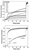
Peptide-induced leakage of Tb3+/DPA from phospholipid vesicles. A: Time course of leakage. Vesicles containing Tb3+ were first diluted into buffer containing an excess of DPA and a baseline fluorescence intensity was established. At 2.5 minutes peptide was added from a concentrated solution to achieve P:L = 1:200. Tb3+/DPA complex formation gives rise to a fluorescence intensity that is measured. At 27.5 minutes, Triton X-100 was added to release all contents from the vesicles. Eight of the ten peptides caused a rapid burst of leakage from the vesicle that was nearly complete within 10 minutes. Two of the peptides caused slower leakage, which was essentially complete by about 30 minutes. B. Time dependence of binding and leakage. In this panel we superimpose the time course of binding of the two unusually slow peptides, measured by tryptophan fluorescence, and the time course of leakage measured at P:L = 1:100.
Figure 7
Figure 7
Requenching assay for determining the mechanism of contents leakage from lipid bilayer vesicles. Various concentrations of the 10 membrane permeabilizing peptides were added to a constant vesicle solution with entrapped dye (ANTS) and quencher (DPX) as described in the text. After leakage had stopped (∼30 min), the requenching assay was used to determine the fraction of ANTS that had been released (fout) and the degree of quenching of the ANTS that remains entrapped within vesicles (Qin). Quenching is defined as a fractional fluorescence and thus ranges from ∼0.5 (initial quenching level in these vesicle preparations) to 1 (unquenched). Theoretical curves for all-or-none and for graded release are shown. These peptides cause predominantly all-or-none release in which a vesicle either releases all of its contents, or none of them.
Figure 8
Figure 8
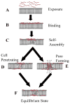
Enhanced mechanistic model for the actions of the pore forming peptides in phospholipid bilayers. We propose that an amended “carpet” or “sinking raft” model of peptide pore formation explains our observations in the following steps: A: Intact membranes are exposed to peptide. B: Peptide binds to the outer monolayer due to hydrophobic and electrostatic interactions. C: Peptides self-assemble into peptide-lipid domains in the outer monolayers. D, E: Relief of the transbilayer asymmetry simultaneously through D: a cell-penetrating peptide pathway that causes little or no leakage and through E: a cooperative pore formation which causes a vesicle to lose all of its entrapped contents. F: the equilibrium state is reached in which the transbilayer asymmetry has been fully dissipated and pore formation no longer occurs.

References

    1. White SH, Wimley WC. Membrane protein folding and stability: physical principles. Annu Rev Biophys Biomol Struct. 1999;28:319–365. - PubMed
    1. White SH, Wimley WC. Hydrophobic interactions of peptides with membrane interfaces. Biochim Biophys Acta. 1998;1376(3):339–352. - PubMed
    1. Nguyen LT, Schibli DJ, Vogel HJ. Structural studies and model membrane interactions of two peptides derived from bovine lactoferricin. J Pept Sci. 2005;11(7):379–389. - PubMed
    1. Kondejewski LH, Jelokhani-Niaraki M, Farmer SW, Lix B, Kay CM, Sykes BD, Hancock RE, Hodges RS. Dissociation of antimicrobial and hemolytic activities in cyclic peptide diastereomers by systematic alterations in amphipathicity. J Biol Chem. 1999;274(19):13181–13192. - PubMed
    1. Shai Y, Oren Z. From “carpet” mechanism to de-novo designed diastereomeric cell-selective antimicrobial peptides. Peptides. 2001;22(10):1629–1641. - PubMed

Publication types