. 2008 Nov;14(11):1821-34.
doi: 10.1089/ten.tea.2007.0222.

Transient exposure to transforming growth factor beta 3 under serum-free conditions enhances the biomechanical and biochemical maturation of tissue-engineered cartilage

Affiliations

Transient exposure to transforming growth factor beta 3 under serum-free conditions enhances the biomechanical and biochemical maturation of tissue-engineered cartilage

Benjamin A Byers et al. Tissue Eng Part A. 2008 Nov.

Abstract

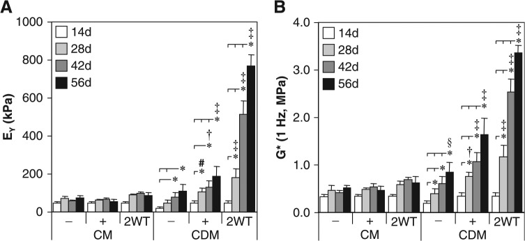
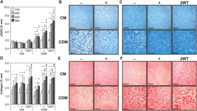
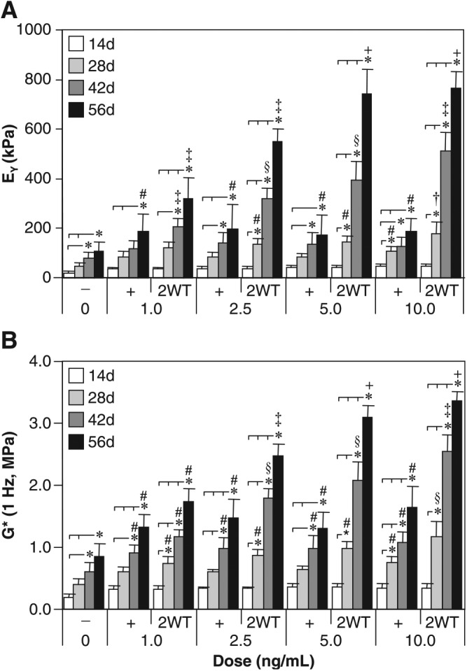
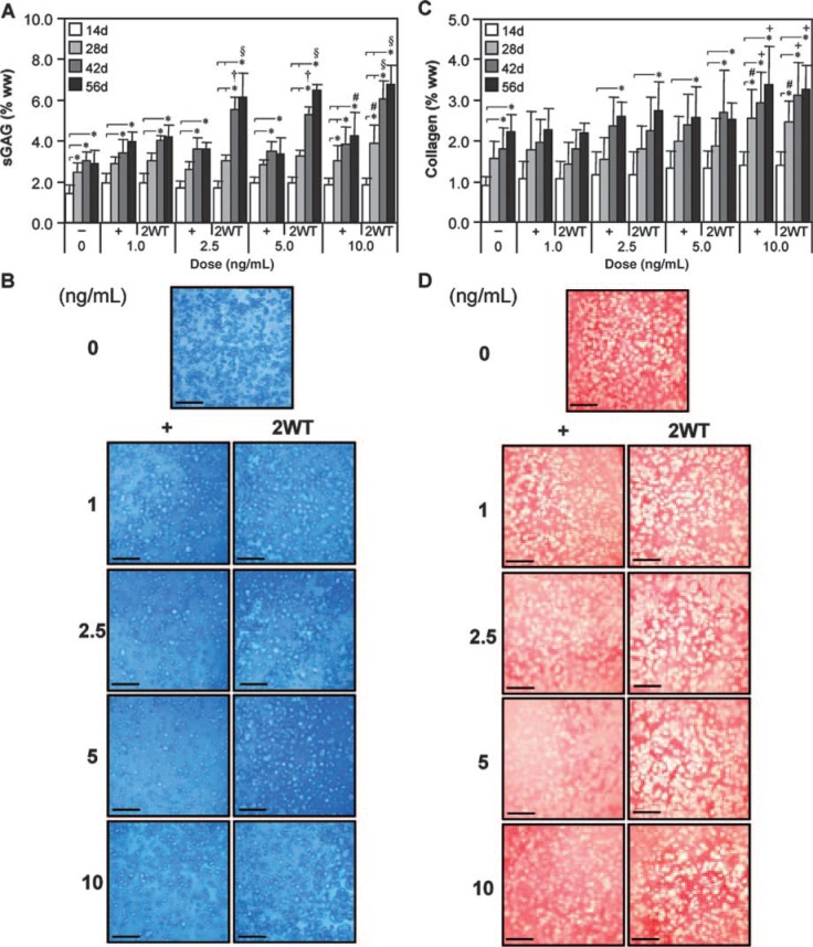
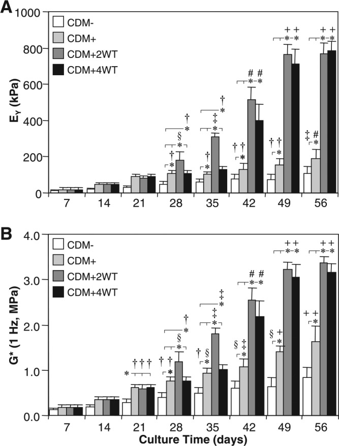
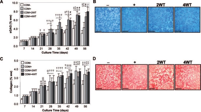
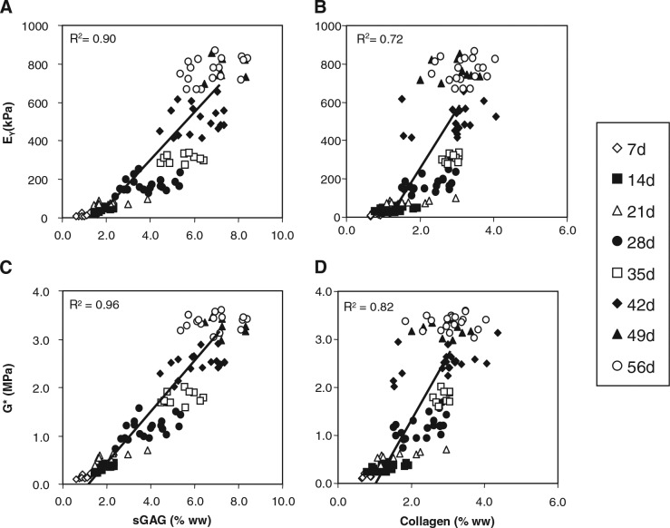
A goal of cartilage tissue engineering is the production of cell-laden constructs possessing sufficient mechanical and biochemical features to enable native tissue function. This study details a systematic characterization of a serum-free (SF) culture methodology employing transient growth factor supplementation to promote robust maturation of tissue-engineered cartilage. Bovine chondrocyte agarose hydrogel constructs were cultured under free-swelling conditions in serum-containing or SF medium supplemented continuously or transiently with varying doses of transforming growth factor beta 3 (TGF-beta3). Constructs were harvested weekly or bi-weekly and assessed for mechanical and biochemical properties. Transient exposure (2 weeks) to low concentrations (2.5-5 ng/mL) of TGF-beta3 in chemically defined medium facilitated robust and highly reproducible construct maturation. Constructs receiving transient TGF-beta3 exposure achieved native tissue levels of compressive modulus (0.8 MPa) and proteoglycan content (6-7% of wet weight) after less than 2 months of in vitro culture. This maturation response was far superior to that observed after continuous growth factor supplementation or transient TGF-beta3 treatment in the presence of serum. These findings represent a significant advance in developing an ex vivo culture methodology to promote production of clinically relevant and mechanically competent tissue-engineered cartilage constructs for implantation to repair damaged articular surfaces.


Figures

FIG. 1.
FIG. 1.
Mechanical properties of cultured chondrocyte-laden constructs as a function of transient transforming growth factor beta 3 (TGF-β3) treatment. Constructs were cultured up to 56 days in serum-containing growth medium (CM) or a chemically defined medium (CDM) without TGF-β3 supplementation (CM– and CDM–), with continuous TGF-β3 (10 ng/mL) exposure (CM + and CDM+), or transient TGF-β3 exposure for the first 2 weeks of culture (CM + 2WT and CDM + 2WT). The (A) equilibrium Young's modulus (EY) and (B) dynamic modulus (G*, 1 Hz) demonstrated robust time-dependent increases after transient TGF-β3 treatment under serum-free conditions (CDM + 2WT), whereas a similar response was not observed in serum-containing medium (CM + 2WT). Data represent means ± standard deviations of 8 to 22 samples from two to five replicate studies, depending on treatment. Statistical differences (p < 0.05) based on pairwise comparisons are depicted within treatment over time and between treatments within time: *between time points within treatment; #versus CDM– within time point; versus CM–, CM + , and CDM– within time point; versus all other treatments within time point; §versus CM– and CM + within time point.
FIG. 2.
FIG. 2.
Biochemical and histological analyses of cultured chondrocyte-laden hydrogel constructs as a function of transient transforming growth factor beta 3 (TGF-β3) treatment. Constructs were cultured up to 56 days in serum-containing growth medium (CM) or a chemically defined medium (CDM) without TGF-β3 supplementation (CM– and CDM–), with continuous TGF-β3 (10 ng/mL) exposure (CM + and CDM+), or transient TGF-β3 exposure for the first 2 weeks of culture (CM + 2WT and CDM + 2WT). (A) Sulfated glycosaminoglycan (sGAG) content. Substantial increases were seen in all serum-free (SF) cultures (CDM–, CDM + , and CDM + 2WT) over time. The most pronounced enhancement was observed in CDM + 2WT cultures, correlating to the observed increase in mechanical properties. (B, C) Histology—Alcian blue. Enhanced staining was seen over time in culture under SF culture conditions. Representative micrographs are depicted at (B) 14 days and (C) 56 days (bar = 500 μm). (D) Collagen content. Time-dependent increases were seen in all culture conditions, although to a much lesser extent than observed for sGAG and at levels well below those of native cartilage. (E, F) Histology—Picrosirius red. Accumulation of a collagenous matrix was detected at (E) 14 days and (F) 56 days (bar = 500 μm). In (A) and (D), data represent the means ± standard deviations of 8 to 22 samples from two to five replicate studies, depending on treatment. Statistical differences (p < 0.05) based on pairwise comparisons are depicted within treatment over time and between treatments within time: *between time points within treatment; +versus CM–, CM + , and CM + 2WT within time point; $versus CM–, CM + , CM + 2WT, and CDM– within time point; versus all other treatments within time point; #versus CDM– within time point. Color images available online at www.liebertonline.com/ten.
FIG. 3.
FIG. 3.
Mechanical properties of cultured chondrocyte-laden constructs as a function of transforming growth factor beta 3 (TGF-β3) dose. Constructs were cultured up to 56 days in chemically defined medium (CDM) without TGF-β3 supplementation (CDM–) or with continuous (CDM+) or 2-week transient (CDM + 2WT) exposure to varying doses of TGF-β3 (1.0, 2.5, 5.0, and 10.0 ng/mL). All groups receiving continuous doses of TGF-β3 demonstrated comparable enhancement of (A) equilibrium Young's moduli (EY) and (B) dynamic moduli (G*, 1 Hz) with time in culture; however, a robust dose-dependence was observed in groups receiving transient TGF-β3 exposure. Maximal response in EY and G* were achieved with a transient dose of at least 5.0 ng/mL. Data represent the means ± standard deviations of 8 to 22 samples from two to five replicate studies, depending on treatment. Statistical differences (p < 0.05) based on pairwise comparisons are depicted within treatment over time and between treatments within time: *between time points within treatment; #versus CDM– within time point; versus all other treatments within time point; versus CDM– and all doses of CDM + within time point; §versus CDM–, all doses of CDM + , and 1 ng/mL of CDM + 2WT within time point; +versus CDM–, all doses of CDM + , and 1.0 and 2.5 ng/mL of CDM + 2WT within time point.
FIG. 4.
FIG. 4.
Biochemical and histological analyses of cultured chondrocyte-laden constructs as a function of transforming growth factor beta 3 (TGF-β3) dose. Constructs were cultured up to 56 days in chemically defined medium (CDM) without TGF-β3 supplementation (CDM−) or with continuous (CDM+) or 2-week transient (CDM + 2WT) exposure to varying doses of TGF-β3 (1.0, 2.5, 5.0, and 10.0 ng/mL). (A) Sulfated glycosaminoglycan (sGAG) content. Transient TGF-β3 exposure resulted in a dose-dependent increase in sGAG content, with peak levels attained at 2.5 ng/mL of CDM + 2WT at 56 days. (B) Histology—Alcian blue. Robust staining indicating sGAG accumulation was seen across all doses of constant and transient TGF-β3 treatment after 56 days of culture (bar = 500 μm). (C) Collagen content. Unlike with sGAG, collagen content increased only moderately over time, with the highest dose providing the most-significant collagen accumulation. (D) Histology—Picrosirius red. Staining at 56 days confirmed accumulation of a collagenous matrix (bar = 500 μm). In (A) and (C), data represent the means ± standard deviations of 8 to 22 samples from two to five replicate studies, depending on treatment. Statistical differences (p < 0.05) based on pairwise comparisons are depicted within treatment over time and between treatments within time: *between time points within treatment; #versus CDM− within time point; versus CDM− and all doses of CDM + within time point; §versus CDM−, all doses of CDM+, and 1 ng/mL of CDM + 2WT within time point; +versus CDM−, 1 ng/mL of CDM + , and 1 ng/mL of CDM + 2WT within time point. Color images available online at www.liebertonline.com/ten.
FIG. 5.
FIG. 5.
Mechanical properties of cultured chondrocyte-laden constructs as a function of transforming growth factor beta 3 (TGF-β3) treatment duration. Constructs were harvested weekly up to 56 days after culture in chemically defined medium (CDM) without TGF-β3 supplementation (CDM–) or with continuous (CDM+) or transient TGF-β3 exposure for the first 2 weeks (CDM + 2WT) or 4 weeks (CDM + 4WT) of culture. Both groups receiving transient growth factor treatment exhibited robust time-dependent increases in (A) equilibrium Young's moduli (EY) and (B) dynamic moduli (G*, 1 Hz) and achieved comparable equilibrium values at 56 days. Data represent the means ± standard deviations of 10 to 22 samples from two to five replicate studies depending on treatment. Statistical differences (p < 0.05) based on pairwise comparisons are depicted within treatment over time and between treatments within time: *between treatments within time point; versus day 14 and all prior time points within treatment; §versus day 21 and all prior time points within treatment; versus day 28 and all prior time points within treatment; #versus day 35 and all prior time points within treatment; +versus day 42 and all prior time points within treatment.
FIG. 6.
FIG. 6.
Biochemical and histological analyses of cultured chondrocyte-laden constructs as a function of transforming growth factor beta 3 (TGF-β3) treatment duration. Constructs were harvested weekly up to 56 days after culture in chemically defined medium (CDM) without TGF-β3 supplementation (CDM–) or with continuous (CDM+) or transient TGF-β3 exposure for the first 2 weeks (CDM + 2WT) or 4 weeks of culture (CDM + 4WT). (A) Sulfated glycosaminoglycan (sGAG) content. All treatment groups exhibited time-dependent increases in sGAG content, with the transient exposure groups attaining the highest levels in correlation to the observed mechanical properties for these culture conditions. (B) Histology—Alcian blue. Robust staining indicating accumulation of sulfated proteoglycan-rich extracellular matrix was seen in all treatments at 56 days (bar = 500 μm). (C) Collagen content. Similar to sGAG content, collagen content demonstrated time-dependent increases for all treatment groups, although transient TGF-β3 exposure did not enhance collagen deposition more than continuous growth factor supplementation. (D) Histology—Picrosirius red. Staining at 56 days demonstrated accumulation of a collagenous matrix (bar = 500 μm). In (A) and (C), data represent the means ± standard deviations of 10 to 22 samples from two to five replicate studies depending on treatment. Statistical differences (p < 0.05) based on pairwise comparisons are depicted within treatment over time and between treatments within time: *between treatments within time point; @versus day 7 within treatment; versus day 14 and all prior time points within treatment; §versus day 21 and all prior time points within treatment; versus day 28 and all prior time points within treatment; #versus day 35 and all prior time points within treatment; +versus day 42 and all prior time points within treatment. Color images available online at www.liebertonline.com/ten.
FIG. 7.
FIG. 7.
Correlation of equilibrium Young's moduli (EY) and dynamic moduli (G*) with sulfated glycosaminoglycan (sGAG) or collagen contents of cultured chondrocyte-laden constructs. (A) EY with sGAG, (B) EY with collagen, (C) G* with sGAG, and (D) G* with collagen. A linear correlation was seen with all parameters in constructs examined from day 7 to day 56 after culture in a chemically defined medium containing transiently supplemented transforming growth factor beta 3 (10 ng/mL) for the first 2 weeks of culture. Coefficient of determination values ranged from 0.72 to 0.96.

References

    1. Ateshian G.A. Hung C.T. Patellofemoral joint biomechanics and tissue engineering. Clin Orthop Relat Res. 2005:81. - PMC - PubMed
    1. Guilak F. Sah R.L. Setton L.A. Physical Regulation of Cartilage Metabolism, in Basic Orthopaedic Biomechanics. In: Mow V.C., editor; Hayes W.C., editor. Lippincott-Raven; Philadelphia: 1997. pp. 179–207.
    1. Vunjak-Novakovic G. Obradovic B. Martin I. Bursac P.M. Langer R. Freed L.E. Dynamic cell seeding of polymer scaffolds for cartilage tissue engineering. Biotechnol Prog. 1998;14:193. - PubMed
    1. Freed L.E. Langer R. Martin I. Pellis N.R. Vunjak-Novakovic G. Tissue engineering of cartilage in space. Proc Natl Acad Sci U S A. 1997;94:13885. - PMC - PubMed
    1. Buschmann M.D. Gluzband Y.A. Grodzinsky A.J. Hunziker E.B. Mechanical compression modulates matrix biosynthesis in chondrocyte/agarose culture. J Cell Sci. 1995;108((Pt 4)):1497. - PubMed

Publication types

LinkOut - more resources