. 2008 Jul;72(1):34-49.
doi: 10.1111/j.1747-0285.2008.00679.x.

Development of broad-spectrum halomethyl ketone inhibitors against coronavirus main protease 3CL(pro)

Affiliations

Development of broad-spectrum halomethyl ketone inhibitors against coronavirus main protease 3CL(pro)

Usman Bacha et al. Chem Biol Drug Des. 2008 Jul.

Abstract

Coronaviruses comprise a large group of RNA viruses with diverse host specificity. The emergence of highly pathogenic strains like the SARS coronavirus (SARS-CoV), and the discovery of two new coronaviruses, NL-63 and HKU1, corroborates the high rate of mutation and recombination that have enabled them to cross species barriers and infect novel hosts. For that reason, the development of broad-spectrum antivirals that are effective against several members of this family is highly desirable. This goal can be accomplished by designing inhibitors against a target, such as the main protease 3CL(pro) (M(pro)), which is highly conserved among all coronaviruses. Here 3CL(pro) derived from the SARS-CoV was used as the primary target to identify a new class of inhibitors containing a halomethyl ketone warhead. The compounds are highly potent against SARS 3CL(pro) with K(i)'s as low as 300 nM. The crystal structure of the complex of one of the compounds with 3CL(pro) indicates that this inhibitor forms a thioether linkage between the halomethyl carbon of the warhead and the catalytic Cys 145. Furthermore, Structure Activity Relationship (SAR) studies of these compounds have led to the identification of a pharmacophore that accurately defines the essential molecular features required for the high affinity.


Figures

Figure 1
Figure 1
The aligned structures of SARS 3CLpro (PDB ID 1Q2W), HCoV 229E 3CLpro (PDB ID 1P9S), IBV 3CLpro (PDB ID 2Q6D), and TGEV 3CLpro (PDB ID 1LVO) are shown in ribbon representation and coloured blue, yellow, red and green respectively. An arrow points to the position of the active site cavity. The Cα of each structure was used in the alignment and all structures were aligned to the SARS 3CLpro structure. The RMSD values from the alignment were 0.889 Å for HCoV 229E 3CLpro (over 247 Cα), 1.302 Å for IBV 3CLpro (over 247 Cα), and 1.8 Å for TGEV 3CLpro (over 272 Cα).
Figure 6
Figure 6
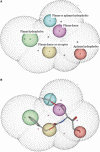
The 3D‐pharmacophore query that was used for virtual screening. (panel A) The features of the pharmacophore are shown as spheres in dot configuration along with the external shell which is shown in grey dot configuration. The aplanar hydrophobic feature is rendered in red, planar hydrophobic feature in green, planar donor feature in purple, planar or aplanar hydrophobic feature in blue and planar donor and acceptor in yellow. (panel B) The 3D‐pharmacophore query aligned with the selected conformation of Compound 4 is shown. The conformation of the selected molecule was dictated by the orientation of the spheres representing each pharmacophore feature. The warhead feature is specified by the aplanar hydrophobic feature (red), P1 residue by the planar donor (purple) and a planar or aplanar hydrophobic feature (blue), compound backbone by a planar donor or acceptor (yellow), and the terminal group is defined by a planar hydrophobic feature (green).
Figure 2
Figure 2
The chemical structure of KNI‐30001. The structures were generated using ChemDraw Ultra 6.0 (Cambridge Software). The compound consists of a trifluoromethyl ketone warhead with a Glu in the P1 position, Leu at P2 and Val at P3.
Figure 3
Figure 3
Calorimetric titration of SARS 3CLpro with Compound 4. In this experiment, each peak represents the injection of 10 μL of 3CLpro (125 μm) into the calorimetric cell (1.4272 mL) containing Compound 4 at a concentration of 8 μm. The experiment was performed at 25 °C in buffer containing 10 mm sodium phosphate, 10 mm NaCl, 1 mm TCEP, 1 mm EDTA, pH 7.4, with a 2% final DMSO concentration.
Figure 4
Figure 4
Location of the Compound 4 binding site. (panel A) Crystal structure of SARS 3CLpro bound to Compound 4 showing the location of the ligand within the SARS protease monomer. The protease is depicted in ribbon representation with the catalytic residues His41 and Cys145 shown as yellow sticks. The ligand is shown in stick representation and is coloured by atom type with oxygen in red, nitrogen in blue, and carbon in green. (Panel B) Key residues involved in the protein–ligand interaction. The backbone of the protein is shown as blue ribbon. The side chains of the protein and ligand are coloured by atom type with oxygen in red, nitrogen in blue, sulfur in tan, and carbon in either green or yellow for the ligand and protein, respectively. (panel C) Orientation of the ligand within the active site cavity of SARS 3CLpro. The ligand is depicted in stick representation and is coloured by atom type as in (panel A) and (panel B). The protein is shown in surface representation with each of the defined substrate subsites highlighted. S1 is shown in red, S2 in rose, and S4 in blue. Cys145 is depicted in yellow. Non‐subsite residues are shown in grey.
Figure 5
Figure 5
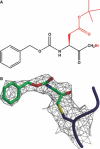
Structure of Compound 4. (panel A) The chemical structure of Compound 4 is shown with the atoms of the ligand observed in the crystal structure coloured in black and the atoms not observed in red. (panel B) The 2Fo−Fc electron density map corresponding to Compound 4 bound to Cys145 is shown, contoured to 1 sigma. The protein and ligand are coloured by atom type with oxygen in red, nitrogen in blue, sulphur in yellow, and carbon in either green or blue for the ligand and protein, respectively.

References

    1. Wat D. (2004) The common cold: a review of the literature. Eur J Intern Med;15:79–88. - PMC - PubMed
    1. Peiris J.S., Lai S.T., Poon L.L., Guan Y., Yam L.Y., Lim W., Nicholls J. et al. (2003) Coronavirus as a possible cause of severe acute respiratory syndrome. Lancet;361:1319–1325. - PMC - PubMed
    1. Rota P.A., Oberste M.S., Monroe S.S., Nix W.A., Campagnoli R., Icenogle J.P., Penaranda S. et al. (2003) Characterization of a novel coronavirus associated with severe acute respiratory syndrome. Science;300:1394–1399. - PubMed
    1. Marra M.A., Jones S.J., Astell C.R., Holt R.A., Brooks‐Wilson A., Butterfield Y.S., Khattra J. et al. (2003) The Genome sequence of the SARS‐associated coronavirus. Science;300:1399–1404. - PubMed
    1. Van Der Hoek L., Pyrc K., Jebbink M.F., Vermeulen‐Oost W., Berkhout R.J., Wolthers K.C., Wertheim‐van Dillen P.M. et al. (2004) Identification of a new human coronavirus. Nat Med;10:368–373. - PMC - PubMed

Publication types

MeSH terms

LinkOut - more resources