. 2008 Jul 8:3:13.
doi: 10.1186/1751-0473-3-13.

Wndchrm - an open source utility for biological image analysis

Affiliations

Wndchrm - an open source utility for biological image analysis

Lior Shamir et al. Source Code Biol Med. .

Abstract

Background: Biological imaging is an emerging field, covering a wide range of applications in biological and clinical research. However, while machinery for automated experimenting and data acquisition has been developing rapidly in the past years, automated image analysis often introduces a bottleneck in high content screening.

Methods: Wndchrm is an open source utility for biological image analysis. The software works by first extracting image content descriptors from the raw image, image transforms, and compound image transforms. Then, the most informative features are selected, and the feature vector of each image is used for classification and similarity measurement.

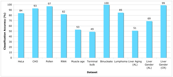
Results: Wndchrm has been tested using several publicly available biological datasets, and provided results which are favorably comparable to the performance of task-specific algorithms developed for these datasets. The simple user interface allows researchers who are not knowledgeable in computer vision methods and have no background in computer programming to apply image analysis to their data.

Conclusion: We suggest that wndchrm can be effectively used for a wide range of biological image analysis tasks. Using wndchrm can allow scientists to perform automated biological image analysis while avoiding the costly challenge of implementing computer vision and pattern recognition algorithms.


Figures

Figure 1
Figure 1
Time required for processing a single image. The time required for computing image features for a single image is linear to the number of pixels for images no larger than 512 × 512 pixels.
Figure 2
Figure 2
An example phylogeny. The phylogeny shows the visual similarities of 16 classes, where each class is a set of 25 images of Drosophila cells with single gene knockdown. The gene classes are specified by their CG numbers. As can be learned from the phylogeny, the resulting phenotypes when knocking down genes CG10895 and CG10873 (which have a substrate-kinase relationship) are very similar to each other, while the phenotypes of gene CG12284 (cell death), CG3733 (unknown function) and untreated cells are different from the other cells.
Figure 3
Figure 3
Classification accuracy using iicbu-2008. As the graph shows, some of the image datasets were classified with very high accuracy, such as Pollen, Binucleate, and Liver age (gender). Other datasets such as HeLa, Lymphoma and RNAi were classified in accuracy of 80–85%, and the datasets Muscle Age, Terminal Bulb and Liver Aging (age) provided classification accuracy of around 50%.
Figure 4
Figure 4
Phylogeny of the worm terminal bulb aging. The phylogeny that was automatically generated by wndchrm shows a class order that is in agreement with the chronological ages.

References

    1. Swedlow JR, Goldberg IG, Brauner E, Sorger PK. Image informatics and quantitative analysis of biological images. Science. 2003;300:100–102. - PMC - PubMed
    1. Goldberg I, Allan C, Burel JM, Creager D, Falconi A, Hochheiser H, Johnston J, Mellen J, Sorger PK, Swedlow JR. Image informatics and quantitative analysis of biological images. Genome Biology. 2005;6:R47. - PMC - PubMed
    1. Swedlow JR. The Open Microscopy Environment: A collaborative data modeling and software development project for biological image informatics. In: Spencer L, Frischknecht F, editor. Imaging Cellular and Molecular Biological Functions. Berlin: Springer; 2007. pp. 71–92.
    1. Carpenter AE, Jones TR, Lamprecht MR, Clarke C, Kang IH, Friman O, Guertin DA, Chang JH, Lindquist RA, Moffat J, Golland P, Sabatini DM. CellProfiler: image analysis software for identifying and quantifying cell phenotypes. Genome Biology. 2006;7:R100. - PMC - PubMed
    1. Orlov N, Shamir L, Macura T, Johnston J, Eckley DM, Goldberg I. WND-CHARM – Multi purpose classification using compound image transforms. Pattern Recognition Letters. - PMC - PubMed

LinkOut - more resources