. 2008 Dec;11(8):1123-34.
doi: 10.1017/S1461145708009024. Epub 2008 Jul 9.

Histone deacetylase inhibitors up-regulate astrocyte GDNF and BDNF gene transcription and protect dopaminergic neurons

Affiliations

Histone deacetylase inhibitors up-regulate astrocyte GDNF and BDNF gene transcription and protect dopaminergic neurons

Xuefei Wu et al. Int J Neuropsychopharmacol. 2008 Dec.

Abstract

Parkinson's disease (PD) is characterized by the selective and progressive loss of dopaminergic (DA) neurons in the midbrain substantia nigra. Currently, available treatment is unable to alter PD progression. Previously, we demonstrated that valproic acid (VPA), a mood stabilizer, anticonvulsant and histone deacetylase (HDAC) inhibitor, increases the expression of glial cell line-derived neurotrophic factor (GDNF) and brain-derived neurotrophic factor (BDNF) in astrocytes to protect DA neurons in midbrain neuron-glia cultures. The present study investigated whether these effects are due to HDAC inhibition and histone acetylation. Here, we show that two additional HDAC inhibitors, sodium butyrate (SB) and trichostatin A (TSA), mimic the survival-promoting and protective effects of VPA on DA neurons in neuron-glia cultures. Similar to VPA, both SB and TSA increased GDNF and BDNF transcripts in astrocytes in a time-dependent manner. Furthermore, marked increases in GDNF promoter activity and promoter-associated histone H3 acetylation were noted in astrocytes treated with all three compounds, where the time-course for acetylation was similar to that for gene transcription. Taken together, our results indicate that HDAC inhibitors up-regulate GDNF and BDNF expression in astrocytes and protect DA neurons, at least in part, through HDAC inhibition. This study indicates that astrocytes may be a critical neuroprotective mechanism of HDAC inhibitors, revealing a novel target for the treatment of psychiatric and neurodegenerative diseases.


Figures
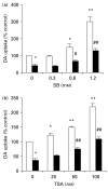
Figure 1
Figure 1
Treatment with histone deacetylase (HDAC) inhibitors preserves dopaminergic (DA) neuronal function in 1-methyl-4-phenylpyridinium (MPP+)-treated neuron-glia cultures. Midbrain neuron-glia cultures were treated with vehicle, sodium butyrate (SB) (a) or trichostatin A (TSA) (b) at the indicated concentrations in the presence (■) or absence (□) of 0.5 μM MPP+. MPP+ was added 30 min after SB and TSA pretreatment. DA neuronal function was evaluated by the [3H]dopamine uptake assay 7 d after the varying treatments. Results are expressed as percent of control and represent means ±S.E. of three separate experiments. * p<0.05, ** p<0.01, compared to untreated control groups; # p<0.05, ## p<0.01, compared to MPP+ alone group.
Figure 2
Figure 2
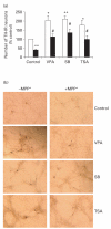
Histone deacetylase (HDAC) inhibitors prevent cell loss of dopaminergic (DA) neurons in 1-methyl-4-phenylpyridinium (MPP+)-treated neuron-glia cultures. Midbrain neuron-glia cultures were treated with vehicle, 1.2 mM valproic acid (VPA), 1.2 mM sodium butyrate (SB), or 100 nM trichostatin A (TSA) in the presence (■) or absence (□) of 0.5 μM MPP+.MPP+ was added 30 min after SB and TSA pretreatment. DA neuronal cell loss was assessed by tyrosine hydroxylase (TH) immunostaining 7 d after treatment (a). Results are expressed as percent of control and are the means±S.E. of three experiments. * p<0.05, ** p<0.01, compared to untreated control groups; # p<0.05, compared to MPP+ alone group. (b) Representative micrographs of morphological changes observed following treatment, as indicated in panel (a).
Figure 3
Figure 3
Histone deacetylase (HDAC) inhibitors enhance glial cell line-derived neurotrophic factor (GDNF) promoter activity. C6 glioma cells were transfected with a pGL3-GDNF-1412/+24 reporter construct and treated with 1.2 mM valproic acid (VPA), 1.2 mM sodium butyrate (SB) or 100 nM trichostatin A (TSA) for 24 h before the GDNF promoter activity was assayed in the luciferase reporter system. Results are the means±S.E. of three independent experiments and expressed as relative luciferase activity (firefly luciferase relative to Renilla luciferase activity) compared to untreated cells. * p<0.05, compared to control groups.
Figure 4
Figure 4
Glial cell line-derived neurotrophic factor (GDNF) promoter-associated histone H3 is hyperacetylated in astrocytes treated with histone deacetylase (HDAC) inhibitors. (a) Schematic representation of the rat GDNF promoter. The locations of GDNF promoter primers are indicated with arrowheads. (b) Enriched cortical astrocyte cultures were treated with 1.2 mM valproic acid (VPA), 1.2 mM sodium butyrate (SB), or 100 nM trichostatin A (TSA) for 5, 24 or 48 h. Chromatin immunoprecipitation (ChIP) assay was performed using an anti-acetyl-histone H3 antibody. The amount of immunoprecipitated (ChIP DNA) and non-immunoprecipitated genome DNA (input DNA) following 24 h treatment was measured by PCR with GDNF Pa, GDNF Pb and GDNF Pc primer sets. PCR products were run on a 2% agarose gel and stained with ethidium bromide. Representative results from gels for each primer set are shown. (c) Real-time PCR was performed with the GDNF Pc primer set to quantify the ChIP DNA and input DNA after 5 h (□), 24 h (■) or 48 h (■) treatment using a relative standard curve method. The values of the ChIP DNA were normalized to the input DNA. Data are expressed as fold-change over the control and are the means±S.E. of three independent experiments. * p<0.05, ** p<0.01, compared to control groups.

References

    1. Armstrong KJ, Niles LP. Induction of GDNF mRNA expression by melatonin in rat C6 glioma cells. Neuroreport. 2002;13:473–475. - PubMed
    1. Beck KD, Valverde J, Alexi T, Poulsen K, Moffat B, Vandlen RA, Rosenthal A, Hefti F. Mesencephalic dopaminergic neurons protected by GDNF from axotomy-induced degeneration in the adult brain. Nature. 1995;373:339–341. - PubMed
    1. Burke RE, Antonelli M, Sulzer D. Glial cell line-derived neurotrophic growth factor inhibits apoptotic death of postnatal substantia nigra dopamine neurons in primary culture. Journal of Neurochemistry. 1998;71:517–525. - PubMed
    1. Canudas AM, Pezzi S, Canals JM, Pallas M, Alberch J. Endogenous brain-derived neurotrophic factor protects dopaminergic nigral neurons against transneuronal degeneration induced by striatal excitotoxic injury. Brain Research. Molecular Brain Research. 2005;134:147–154. - PubMed
    1. Castro LM, Gallant M, Niles LP. Novel targets for valproic acid: up-regulation of melatonin receptors and neurotrophic factors in C6 glioma cells. Journal of Neurochemistry. 2005;95:1227–1236. - PubMed

Publication types

MeSH terms