. 2008 Aug;36(13):4498-509.
doi: 10.1093/nar/gkn414. Epub 2008 Jul 8.

TAp73beta and DNp73beta activate the expression of the pro-survival caspase-2S

Affiliations

TAp73beta and DNp73beta activate the expression of the pro-survival caspase-2S

Wen Hong Toh et al. Nucleic Acids Res. 2008 Aug.

Abstract

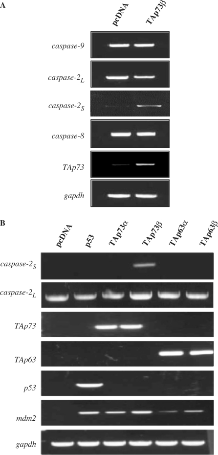
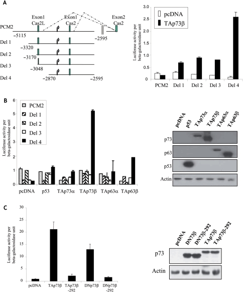
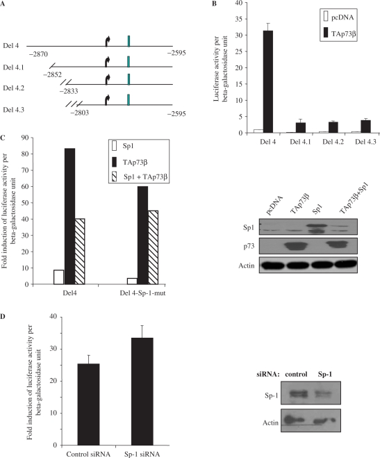
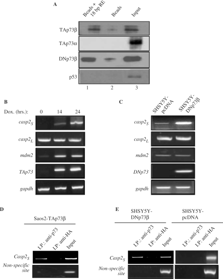
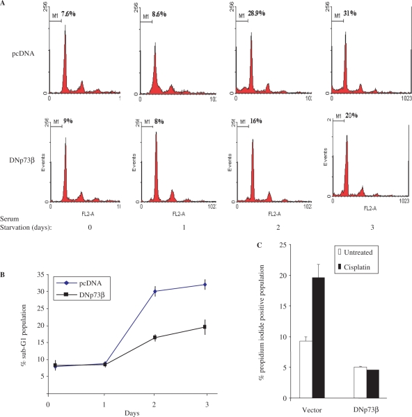
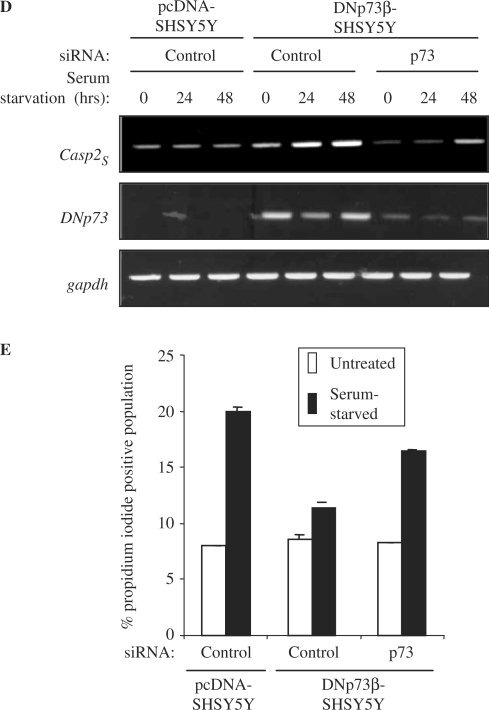
p73, the p53 homologue, exists as a transactivation-domain-proficient TAp73 or deficient deltaN(DN)p73 form. Expectedly, the oncogenic DNp73 that is capable of inactivating both TAp73 and p53 function, is over-expressed in cancers. However, the role of TAp73, which exhibits tumour-suppressive properties in gain or loss of function models, in human cancers where it is hyper-expressed is unclear. We demonstrate here that both TAp73 and DNp73 are able to specifically transactivate the expression of the anti-apoptotic member of the caspase family, caspase-2(S). Neither p53 nor TAp63 has this property, and only the p73beta form, but not the p73alpha form, has this competency. Caspase-2 promoter analysis revealed that a non-canonical, 18 bp GC-rich Sp-1-binding site-containing region is essential for p73beta-mediated activation. However, mutating the Sp-1-binding site or silencing Sp-1 expression did not affect p73beta's transactivation ability. In vitro DNA binding and in vivo chromatin immunoprecipitation assays indicated that p73beta is capable of directly binding to this region, and consistently, DNA binding p73 mutant was unable to transactivate caspase-2(S). Finally, DNp73beta over-expression in neuroblastoma cells led to resistance to cell death, and concomitantly to elevated levels of caspase-2(S.) Silencing p73 expression in these cells led to reduction of caspase-2(S) expression and increased cell death. Together, the data identifies caspase-2(S) as a novel transcriptional target common to both TAp73 and DNp73, and raises the possibility that TAp73 may be over-expressed in cancers to promote survival.


Figures

Figure 1.
Figure 1.
TAp73β, but not p53, TAp73α, TAp63α or TAp63β, induce caspase-2S expression. (A) TAp73β expression induces the up-regulation of caspase-2S but not other caspase transcripts. The pcDNA and TAp73 expression constructs were transfected into H1299 cells for 36 h before RNA extraction and cDNA synthesis. Semi-quantitative RT–PCR was performed for caspase-2L, caspase-8, caspase-9 and caspase-2S transcripts. (B) The pcDNA, p53, TAp63α, TAp63β, TAp73α and TAp73β expression constructs were transfected into H1299 cells similarly and expression of caspase-2L and caspase-2S transcripts were analysed. Mdm2 was used as a positive control and the expression of the transfected p53 family members is shown.
Figure 2.
Figure 2.
Activation of caspase-2S promoter by TAp73β and DNp73β. (A) Left panel shows the schematic of the caspase-2S promoter-luciferase constructs used in the study. Curved arrows represent the TATA box. Exon 1 of caspase-2S and caspase-2L are shown. These constructs were transfected together with TAp73β and β-gal plasmid into H1299 cells for 24 h. The cultures were then lysed and used for luciferase assays (right panel). (B) Activation of Del 4 reporter construct is specific only to TAp73β. The indicated reporter constructs were transfected together with either of the following: p53, TAp63α, TAp63β, TAp73α and TAp73β, together with the β-gal plasmid into H1299 cells for 24 h, prior to analysis of luciferase activity (left panel). Right panel shows immunoblot analysis of the expression of the transfected plasmids. (C) DNA-binding ability of p73β is essential for induction of Del4 promoter. Del4 reporter constructs were transfected together with either TAp73β or TAp73β-292, or DNp73β or DNp73β-292 together with β-gal plasmid into H1299 cells, prior to analysis of luciferase activity (left panel). Right panel shows immunoblot analysis of the expression of the transfected plasmids. All luciferase assays were repeated at least thrice, each time in duplicates. The graphs are representation of average ± SED.
Figure 3.
Figure 3.
Characterization of DNA elements required for activation of caspase-2S promoter by p73. (A) Schematic shows the deletion constructs made sequentially using Del 4. (B) The sequentially truncated Del4 constructs were transfected together with TAp73β and β-gal into H1299 cells for 24 h, prior to luciferase analysis. (C) Del4 and Del4-Sp-1 mutant reporter constructs were transfected together with TAp73β or Sp-1, alone or in combination, into H1299 cells for luciferase analysis. The fold induction of luciferase activity was derived by dividing the values obtained with the respective constructs with that of pcDNA-transfected samples. Right panel shows immunoblot analysis of the expression of the transfected plasmids. (D) Knockdown of Sp-1 does not significantly affect the activation of Del4 promoter by TAp73β. Control or Sp-1 siRNA were transfected into H1299 for 24 h prior to transfection of the Del4, TAp73β and β-gal constructs for luciferase analysis. Fold induction of luciferase activity is shown. Right panel shows efficiency of Sp-1 knockdown by immunoblotting. All luciferase assays were repeated at least thrice, each time in duplicates. The graphs are representation of average ± SED.
Figure 4.
Figure 4.
The p73 binds to the 18-bp sequence element on the caspase-2S promoter in vitro and in vivo. (A) In vitro DNA-GSTp73 binding assay. Purified GST-TAp73α GST-TAp73β GST-DNp73β or GST-p53 proteins were incubated with biotinylated 18 bp caspase-2S promoter containing sequence elements before further incubation with avidin-conjugated beads. GST-p73/p53 without biotinylated DNA but with beads alone was used as negative controls. The beads were washed and separated onto SDS–acrylamide gel for immunoblot analysis with the indicated antibodies. Lane1: GST-protein + 18-bp element + beads. Lane 2: GST-protein + beads. Lane 3: GST-protein lysate. (B–C) Up-regulation of caspase-2S in Saos2-TAp73β inducible cell line (B) and in SH-SY5Y cells stably expressing DNp73β (C). TAp73β was induced by doxocycline (Dox) addition for 14–24 h prior to RNA extraction. RT–PCR was performed to assess expression of caspase-2S, caspase-2L, p73 and mdm2 in both the cell systems. (D and E) ChIP analysis was performed with anti-p73 and anti-HA antibodies using the two cellular systems described earlier. Cells were collected 15 h after TAp73 induction (D). The promoter sequence encompassing the 18-bp elements on the caspase-2S promoter was analysed by PCR (Casp2S). A non-specific site on the caspase-2S promoter was used as negative control. All experiments were repeated at least thrice independently.
Figure 5.
Figure 5.
DNp73β over-expressing SH-SY5Y cells are resistant to cell death. (A and B) SH-SY5Y cells stably over-expressing pcDNA or DNp73β were seeded in 6-well plates in triplicates and serum starved in serum free DMEM for up to 72 h. Cultures were harvested at days 0, 1, 2 and 3 during serum starvation for cell cycle analysis. Representative flow cytometric graphics (A). Average of the sub-G1 population, representing apoptotic cells, for each cell line and time point is plotted ± SED (B). (C) These cells were treated with 20 μM cisplatin for 24 h prior to analysis of total cell death by propidium iodide exclusion assay. (D and E) Knockdown of p73 results in reduced caspase-2S expression and increased cell death. The above cells were transfected with control or p73-siRNA and serum-starved for the indicated time periods. The mRNA analysis was performed to determine expression of caspase-2S, DNp73 and gapdh (D), and cells were harvested after 48 h for analysis of cell death by propidium iodide exclusion assay (E). All experiments were repeated at least thrice independently.
Figure 5.
Figure 5.
DNp73β over-expressing SH-SY5Y cells are resistant to cell death. (A and B) SH-SY5Y cells stably over-expressing pcDNA or DNp73β were seeded in 6-well plates in triplicates and serum starved in serum free DMEM for up to 72 h. Cultures were harvested at days 0, 1, 2 and 3 during serum starvation for cell cycle analysis. Representative flow cytometric graphics (A). Average of the sub-G1 population, representing apoptotic cells, for each cell line and time point is plotted ± SED (B). (C) These cells were treated with 20 μM cisplatin for 24 h prior to analysis of total cell death by propidium iodide exclusion assay. (D and E) Knockdown of p73 results in reduced caspase-2S expression and increased cell death. The above cells were transfected with control or p73-siRNA and serum-starved for the indicated time periods. The mRNA analysis was performed to determine expression of caspase-2S, DNp73 and gapdh (D), and cells were harvested after 48 h for analysis of cell death by propidium iodide exclusion assay (E). All experiments were repeated at least thrice independently.

References

    1. Jost CA, Marin MC, Kaelin W.G., Jr p73 is a simian [correction of human] p53-related protein that can induce apoptosis. Nature. 1997;389:191–194. - PubMed
    1. Kaghad M, Bonnet H, Yang A, Creancier L, Biscan JC, Valent A, Minty A, Chalon P, Lelias JM, Dumont X, et al. Monoallelically expressed gene related to p53 at 1p36, a region frequently deleted in neuroblastoma and other human cancers. Cell. 1997;90:809–819. - PubMed
    1. Stiewe T, Zimmermann S, Frilling A, Esche H, Putzer BM. Transactivation-deficient DeltaTA-p73 acts as an oncogene. Cancer Res. 2002;62:3598–3602. - PubMed
    1. Stiewe T, Theseling CC, Putzer BM. Transactivation-deficient Delta TA-p73 inhibits p53 by direct competition for DNA binding: implications for tumorigenesis. J. Biol. Chem. 2002;277:14177–14185. - PubMed
    1. Liu G, Nozell S, Xiao H, Chen X. DeltaNp73beta is active in transactivation and growth suppression. Mol. Cell Biol. 2004;24:487–501. - PMC - PubMed

Publication types

MeSH terms