Review
. 2008 Aug;9(8):639-49.
doi: 10.1038/nrm2447. Epub 2008 Jul 9.

Shaping cups into phagosomes and macropinosomes

Affiliations
Review

Shaping cups into phagosomes and macropinosomes

Joel A Swanson. Nat Rev Mol Cell Biol. 2008 Aug.

Abstract

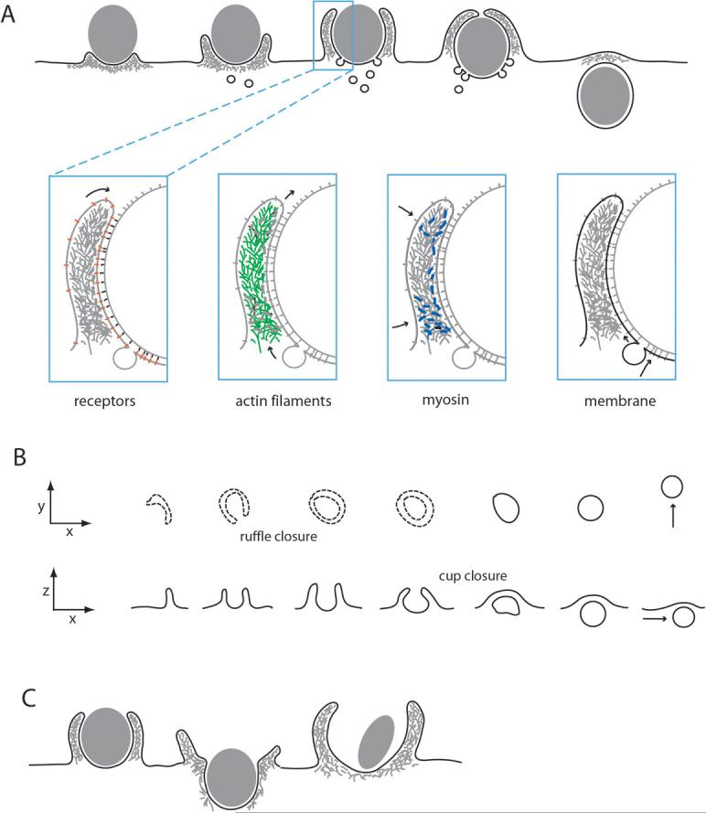
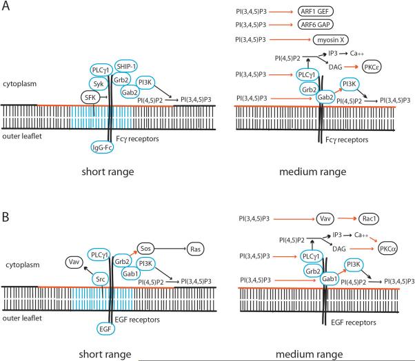
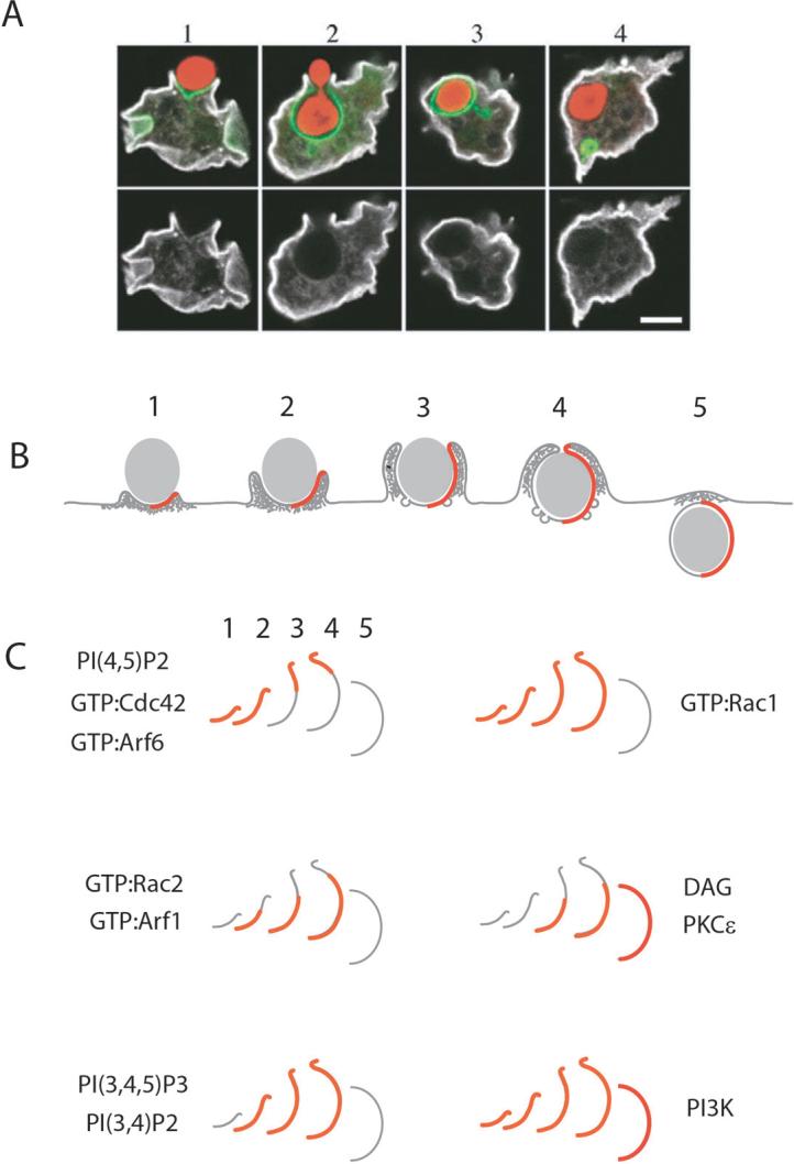
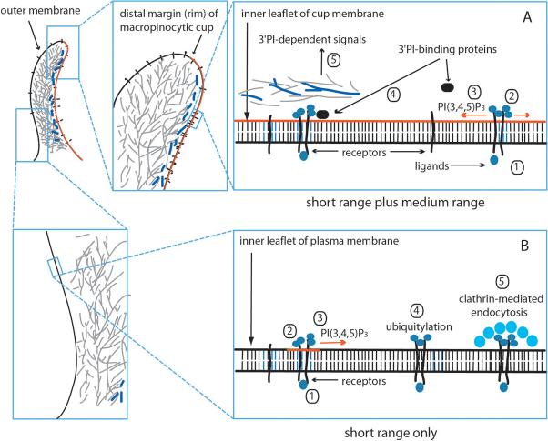
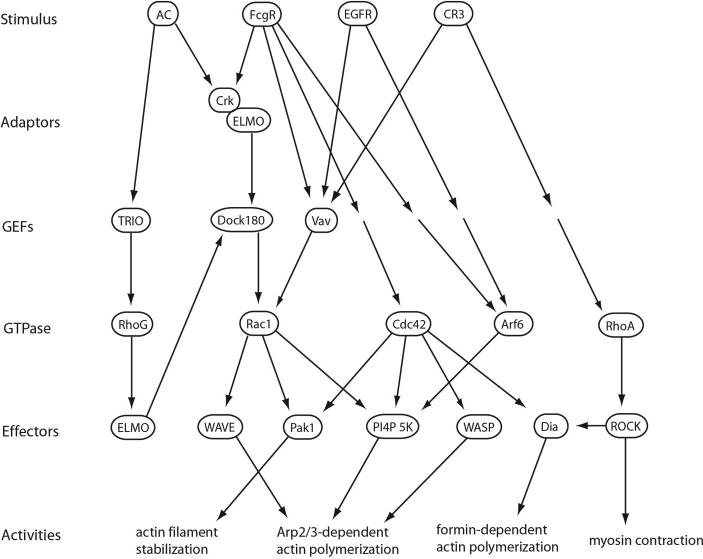
The ingestion of particles or cells by phagocytosis and of fluids by macropinocytosis requires the formation of large endocytic vacuolar compartments inside cells by the organized movements of membranes and the actin cytoskeleton. Fc-receptor-mediated phagocytosis is guided by the zipper-like progression of local, receptor-initiated responses that conform to particle geometry. By contrast, macropinosomes and some phagosomes form with little or no guidance from receptors. The common organizing structure is a cup-shaped invagination of the plasma membrane that becomes the phagosome or macropinosome. Recent studies, focusing on the physical properties of forming cups, indicate that a feedback mechanism regulates the signal transduction of phagocytosis and macropinocytosis.


Figures

Figure 1
Figure 1. The movements of phagocytosis and macropinocytosis
(A) During FcR-mediated phagocytosis, plasma membrane extends over particles as cup-shaped extensions of the cell surface, through progressive interactions of FcR (red) with particle-bound IgG (black). Actin filaments (green) and myosin (blue) are concentrated in the advancing cup, and membrane (black) from intracellular compartments is inserted into the base of the forming cup. Arrows indicate the net direction of receptor movement (receptors), the net displacement of actin filaments by polymerization, depolymerization and contraction (actin), the contraction of the actin-myosin network (myosin) and the net flow of membrane into cups (membrane). (B) Macropinosomes form from cell surface ruffles that close first into open cups (ruffle closure) and then into discrete intracellular vesicles (cup closure). Two aspects of macropinosome formation are presented: the x-y projection indicates the âtop downâ view typically seen in the light microscope and the x-z projection shows a side-view of membrane movements. Dotted lines indicate folds in the plasma membrane. Ruffle closure is the formation of a circular, open cup of plasma membrane. Cup closure is the separation of the macropinosome from the plasma membrane. Arrows indicate macropinosome displacement through cytoplasm. (C) Distinct movements of membranes and actin during various kinds of phagosome formation. (left) Extended, close-fitting cups are typical of the zipper model of FcR-mediated phagocytosis. (middle) During CR3-mediated phagocytosis, phagosomes appear to sink into cytoplasm, although ruffles may accompany the process. (right) In triggered phagocytosis, bacteria are internalized by stimulation of macropinocytosis and recruitment into forming macropinosomes.
Figure 2
Figure 2. Short- and medium-range signaling by activated FcR and EGF receptors
The left panels show dimerized receptors and the proteins that bind (blue circles) or interact (black circles) with those receptors upon ligand-binding. The right panels show medium-range signals that are responsive to receptor-generated PI(3,4,5)P3 (red line in the inner leaflet of the bilayer). Black arrows indicate catalytic activation. Red arrows indicate allosteric activation. (A) FcÎ receptors dimerize in response to ligand binding, leading to conformational changes that favor phosphorylation by Src-family kinases (SFK). Lipid microdomains in the plasma membrane (blue lines) facilitate SFK recruitment to FcR. SFK phosphorylation increases binding and activity of the tyrosine kinase Syk, which stimulates recruitment of PI3K, PLCÎ1, Grb2 and Gab2. Gab2 recruits and activates PI3K in a PI(3,4,5)P3-dependent manner. Grb2 activates the Ras GEF Sos, and also binds the lipid phosphatase SHIP-1, which negatively affects PI(3,4,5)P3 signaling. PI(3,4,5)P3 also activates downstream activities including PLCÎ1 and myosin X. (B) Signaling by EGF receptor dimers is activated by ligand-dependent phosphorylation by Src, which also activates Rac1 via phosphorylation of the GEF Vav. Phosphorylated receptors recruit Grb2, which stimulates activation of the Ras GEF Sos, and recruits Gab1, which activates PI3K. PI(3,4,5)P3 generated by PI3K can activate Gab1, providing a positive-feedback amplification of signals.
Figure 3
Figure 3. Distinct patterns of signaling in phagocytic cups
(A) Phagocytic cups in Dictyostelium discoideum show selective depletion of plasma membrane proteins. The panels show confocal fluorescence microscopy of fluorescent protein chimeras at various stages of phagosome formation. The top row shows distributions of GFP-CRAC (green), which reports the distributions of PI(3,4,5)P3, during phagocytosis of yeast cells (red) Both rows show the distribution of the plasma membrane protein H36 (white), which is selectively depleted from membranes of forming cups. (B) Signals for phagosome formation display distinct patterns during phagosome morphogenesis. The stages of phagocytic cup initiation (1), cup extension (2, 3), closure (4) and separation from the plasma membrane (5) are displayed as patterns in half-cups (red lines). (C) The membranes of half-cups at various stages of phagocytosis are indicated by gray lines. Signal distributions are overlaid as red lines. Early signals include increased concentrations of PI(4,5)P2 and activated Cdc42 and Arf6, which localize to advancing edges of cups. Rac1 is activated early and persists until just after cup closure. Late stages of signaling include activation of Rac2 and Arf1, the generation of DAG and the recruitment of PKCÎ, which are delayed relative to the advance of the cup over the particle. PI3K localizes to forming phagosomes, and its products PI(3,4,5)P3 and PI(3,4)P2 increase during cup formation.
Figure 4
Figure 4. Context-dependent signal transduction in cups
Signaling in the membrane lining macropinocytic or phagocytic cups (red line) (A) is distinct from that in contiguous plasma membrane outside of the cup domain (B). (A) In the cup domain, growth factor or IgG binding to receptors (1) stimulates assembly of receptor complexes (short-range organization) (2). PI3K recruited to receptor complexes stimulates synthesis of PI(3,4,5)P3 (3)(red line), whose concentrations in the inner leaflet of the cup domain increase due to limited diffusion out of the cup and/or positive feedback amplification of its synthesis. Suprathreshold levels of PI(3,4,5)P3 facilitate recruitment or activation of 3â PI-binding proteins (4), which initiate late-stage signals associated with shaping or modifying the cup membrane (5). Such activities are enhanced in cup membranes, possibly due to barriers to diffusion across the distal margin of the cup. (B) Outside of the cup domain, receptor ligation (1) stimulates assembly of a receptor complex (2). PI3K is recruited and synthesizes PI(3,4,5)P3 (3)(red line). However, diffusion or the absence of a positive feedback amplification do not allow PI(3,4,5)P3 concentrations to reach levels that activate medium-range signals. Subsequent ubiquitylation of receptors (4) leads to their down-regulation by clathrin-mediated endocytosis (5).
None

References

    1. Stuart LM, Ezekowitz RA. Phagocytosis and comparative innate immunity: learning on the fly. Nat Rev Immunol. 2008;8:131–141. - PubMed
    1. Watts C, Amigorena S. Antigen traffic pathways in dendritic cells. Traffic. 2000;1:312–317. - PubMed
    1. Blander JM, Medzhitov R. On regulation of phagosome maturation and antigen presentation. Nat Immunol. 2006;7:1029–1035. - PubMed
    1. Reddien PW, Horvitz HR. The engulfment process of programmed cell death in caenorhabditis elegans. Annu Rev Cell Dev Biol. 2004;20:193–221. - PubMed
    1. Amstutz B, et al. Subversion of CtBP1-controlled macropinocytosis by human adenovirus serotype 3. EMBO J. 2008;27:956–969. - PMC - PubMed

Highlighted References

    1. Park, et al. This paper identified a receptor phosphatidylserine presented on apoptotic cells and demonstrated a direct connection to Rac activation via ELMO and Dock180. 2007.
    1. Hall, et al. Demonstrated that the Rac GEF Vav was necessary for CR3-mediated phagocytosis but not for FcR-mediated phagocytosis. This finding is at odds with studies using other cells, which indicated a role for Vav-activated Rac in FcR but not CR3 phagocytosis. 2006.
    1. Amyere, et al. Demonstrated a role for PLCÎ downstream of PI3K during constitutive macropinocytosis in transformed cells. 2000.
    1. Botelho, et al. First demonstration of localized changes in phosphoinositides and diacylglycerol concentrations in unclosed phagocytic cups. 2000.
    1. Mercanti, et al. Demonstrated exclusion of membrane proteins from phagocytic and macropinocytic cups in Dictyostelium discoideum. 2006.

Publication types