. 2008 Jul 9;3(7):e2599.
doi: 10.1371/journal.pone.0002599.

Increased level of extracellular ATP at tumor sites: in vivo imaging with plasma membrane luciferase

Affiliations

Increased level of extracellular ATP at tumor sites: in vivo imaging with plasma membrane luciferase

Patrizia Pellegatti et al. PLoS One. .

Abstract

Background: There is growing awareness that tumour cells build up a "self-advantageous" microenvironment that reduces effectiveness of anti-tumour immune response. While many different immunosuppressive mechanisms are likely to come into play, recent evidence suggests that extracellular adenosine acting at A2A receptors may have a major role in down-modulating the immune response as cancerous tissues contain elevated levels of adenosine and adenosine break-down products. While there is no doubt that all cells possess plasma membrane adenosine transporters that mediate adenosine uptake and may also allow its release, it is now clear that most of extracellularly-generated adenosine originates from the catabolism of extracellular ATP.

Methodology/principal findings: Measurement of extracellular ATP is generally performed in cell supernatants by HPLC or soluble luciferin-luciferase assay, thus it generally turns out to be laborious and inaccurate. We have engineered a chimeric plasma membrane-targeted luciferase that allows in vivo real-time imaging of extracellular ATP. With this novel probe we have measured the ATP concentration within the tumour microenvironment of several experimentally-induced tumours.

Conclusions/significance: Our results show that ATP in the tumour interstitium is in the hundreds micromolar range, while it is basically undetectable in healthy tissues. Here we show that a chimeric plasma membrane-targeted luciferase allows in vivo detection of high extracellular ATP concentration at tumour sites. On the contrary, tumour-free tissues show undetectable extracellular ATP levels. Extracellular ATP may be crucial for the tumour not only as a stimulus for growth but also as a source of an immunosuppressive agent such as adenosine. Our approach offers a new tool for the investigation of the biochemical composition of tumour milieu and for development of novel therapies based on the modulation of extracellular purine-based signalling.


Conflict of interest statement

Competing Interests: FDV serves as a consultant for Duska Scientific Co. (USA) and Affectis Pharmaceuticals AG (Germany), companies involved in the development of P2 receptor-based drugs. A patent is pending on the pmeLUC probe.

Figures

Figure 1
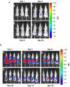
Figure 1. Imaging of healthy nude mice i.v. injected with HEK293-pmeLUC cells.
Mice were injected i.v. with 2×10⁶ HEK293-pmeLUC cells. D-luciferin was administred intraperitoneally. Images were captured 15 minutes after D-luciferin administration. Acquisition time was 3 min, and images were acquired over 2 months every 2 days both from the dorsal (A) and the ventral (B) view. The earliest time point was at 15 min after D-luciferin injection. Only images taken after 1, 3, 15 and 42 days are shown.
Figure 2
Figure 2. Imaging of tumour-bearing nude mice injected with HEK293-pmeLUC cells.
(A) Healthy nude mice were injected i.p. with 2×10⁶ HEK293-pmeLUC cells and monitored for 3 months (only 4 time points are shown). (B) Nude mice were injected i.p. with the human ovarian carcinoma cell line OVCAR-3 (1.5×10⁶). Twenty days post-implantation, HEK293-pmeLUC cells (2×10⁶) were i.p. injected, followed by D-luciferin. OVCAR-3-bearing mice were monitored over 16 days, and were then euthanized according to bioethical regulations.
Figure 3
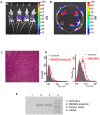
Figure 3. HEK293-pmeLUC cells localize to tumour foci.
(A) Mice were euthanized, the peritoneal cavity was opened and imaged. (B) Single tumour foci were excised and luminescence recorded. (C) Tissue section from one of the tumour foci stained with haematoxylin/eosin. (D) Cytofluorimetric analysis of cells isolated from tumour foci. Staining was performed with monoclonal FITC-labeled rabbit anti-luciferase (red trace on the left panel) or anti-CD51 (red trace on the right panel) Abs. Irrelevant IgG1 Ab (grey trace on both the right and left panel) was used as control. (E) Expression of pmeLUC mRNA in tumour foci excised from mice injected with OVCAR-3 and HEK293-pmeLUC cells. OVCAR-3 and HEK293-pmeLUC cells grown in culture are shown as controls. RT-PCR was performed as described in Materials and Methods.
Figure 4
Figure 4. Comparison of luminescence emission from HEK293-pmeLUC and HEK293-cytLUC cells injected into MZ2-MEL melanoma and healthy tissue.
Nude mice were inoculated with MZ2-MEL cells in the right dorsal hip. After about 20 days, when the tumour was about 1–1.5×1–1.5 cm size, HEK293-pmeLUC (A) or HEK293-cytLUC (B) cells were injected s.c. into the tumour bearing (right) or healthy (left) site.
Figure 5
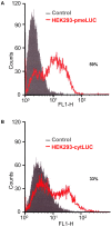
Figure 5. Luciferase expression by HEK293-pmeLUC and HEK293-cytLUC.
Plasma membrane (A) or cytosolic (B) luciferase expression was analyzed with a specific anti-luciferase Ab (see Figure 3). An isotype-matched control mAb was used as control. Luciferase expression is reported as percentage of positive cells.
Figure 6
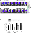
Figure 6. Sensitivity of HEK293-pmeLUC cells to apyrase.
A melanoma was induced as described in Figure 4, and inoculated with HEK293-pmeLUC cells 20 days after implant. Apyrase (20 U in 40 µl of serum free DMEM ) or vehicle (serum free DMEM) was injected into the tumour four days later. Bioluminescence was recorded as indicated. (A) Apyrase-injected, or (B) vehicle-injected mice. (C) Quantitative analysis of light emission from region-of-interest. Data are averages of measurements from 4 mice (n = 4). * p<0.05 (paired t-test).
Figure 7
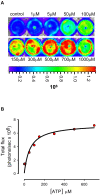
Figure 7. In vitro calibration of HEK293-pmeLUC bioluminescence.
(A) HEK293-pmeLUC cells, 7×10⁴ per well, were incubated in DMEM medium and challenged with increasing concentrations of extracellular ATP and luminescence acquired with the IVIS luminometer for 1 min. In (B) luminescence (total flux expressed as photon per sec) was expressed as a function of the ATP concentration. Control well contained only DMEM medium. D-luciferin was added to all wells at a concentration of 60 µg/ml.

References

    1. Beigi R, Kobatake E, Aizawa M, Dubyak GR. Detection of local ATP release from activated platelets using cell surface-attached firefly luciferase. Am J Physiol Cell Physiol. 1999;276:267–278. - PubMed
    1. Gourine AV, Llaudet E, Dale N, Spyer KM. ATP is a mediator of chemosensory transduction in the central nervous system. Nature. 2005;436:108–111. - PubMed
    1. Gourine AV, Dale N, Llaudet E, Poputnikov DM, Spyer KM, et al. Release of ATP in the central nervous system during systemic inflammation: real-time measurement in the hypothalamus of conscious rabbits. J Physiol. 2007;585:305–316. - PMC - PubMed
    1. Gallina G, Dolcetti L, Serafini P, De Santo C, Marigo I, et al. Tumors induce a subset of inflammatory monocytes with immunosuppressive activity on CD8+ T cells. J Clin Invest. 2006;116:2777–2790. - PMC - PubMed
    1. Albini A, Sporn MB. The tumour microenvironment as a target for chemoprevention. Nat Rev Cancer. 2007;7:139–147. - PubMed

Publication types