. 2008 Jul 9;3(7):e2582.
doi: 10.1371/journal.pone.0002582.

RickA expression is not sufficient to promote actin-based motility of Rickettsia raoultii

Affiliations

RickA expression is not sufficient to promote actin-based motility of Rickettsia raoultii

Premanand Balraj et al. PLoS One. .

Abstract

Background: Rickettsia raoultii is a novel Rickettsia species recently isolated from Dermacentor ticks and classified within the spotted fever group (SFG). The inability of R. raoultii to spread within L929 cells suggests that this bacterium is unable to polymerize host cell actin, a property exhibited by all SFG rickettsiae except R. peacocki. This result led us to investigate if RickA, the protein thought to generate actin nucleation, was expressed within this rickettsia species.

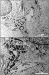
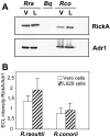
Methodology/principal findings: Amplification and sequencing of R. raoultii rickA showed that this gene encoded a putative 565 amino acid protein highly homologous to those found in other rickettsiae. Using immunofluorescence assays, we determined that the motility pattern (i.e. microcolonies or cell-to-cell spreading) of R. raoultii was different depending on the host cell line in which the bacteria replicated. In contrast, under the same experimental conditions, R. conorii shares the same phenotype both in L929 and in Vero cells. Transmission electron microscopy analysis of infected cells showed that non-motile bacteria were free in the cytosol instead of enclosed in a vacuole. Moreover, western-blot analysis demonstrated that the defect of R. raoultii actin-based motility within L929 cells was not related to lower expression of RickA.

Conclusion/significance: These results, together with previously published data about R. typhi, strongly suggest that another factor, apart from RickA, may be involved with be responsible for actin-based motility in bacteria from the Rickettsia genus.


Conflict of interest statement

Competing Interests: The authors have declared that no competing interests exist.

Figures

Figure 1
Figure 1. (A) Phylogenetic tree of the bacteria belonging to the genus Rickettsia, inferred from a comparison of RickA protein sequences.
This phylogenetic tree was constructed with the NJ method, and using the following RickA protein sequences: R. conorii (rco_ORF0822), R. rickettsii (gi|157828764|), R. slovaca (RISLO1054), R. africae (raf_ORF0824) R. sibirica (ZP00142939), R. massiliae (gi|157844512|), R. felis (gi|67458763|), R. akari (gi|157825980|), R. canadensis (gi|157803553|), R. bellii (gi|91205670|), R. montanensis (gi|75487919|) and R. raoultii (this study). Bootstrap values are indicated at branch nodes. (B) Schematic representation and multiple sequence alignment of the rickettsial RickA proteins from the SFG rickettsiae. G: G-actin binding site. WH2, C and A: WASP-homology 2, central and acidic domains of the WASP-family protein. The numbers of proline-rich repeats (PRR) are indicated in parentheses. Abbrevations are as follows: Rco (R. conorii), Raf (R. africae), Rsl (R. slovaca), Rsi (R. sibirica), Rri (R. rickettsii), Rmo (R. montanensis), Rma (R. massiliae), Rra (R. raoultii), Rfe (R. felis), Rak (R. akari) and Rca (R. canadensis).
Figure 2
Figure 2. Actin tail phenotypes of R. raoultii and R. conorii in L929 cells.
Following a 24 h infection, the bacteria were stained by indirect immunofluorescence using a polyclonal anti-SFG antibody followed by an anti-mouse-Alexa 594 Ig as a secondary antibody (red). F-actin was stained with FITC-phalloidin (green) and nucleic acids were stained with DAPI (blue). Examination of slides with a Leica DM2500 Upright fluorescent microscope (magnifications 40× and 100×), showed that R. raoultii formed microcolonies and multiplied at the center of the cell while R. conorii exhibited actin tails at one pole of the bacterium. The right panel corresponds to a higher-magnification image of R. raoultii microcolony observed with a Leica SPE upright confocal microscope.
Figure 3
Figure 3. TEM of L929 cells infected with R. raoultii.
(A) TEM performed on R. raoultii cultured for 48 h in L929 cells confirmed that bacteria multiply without infecting neighboring cells. (B) A higher magnification showed free bacteria in the cytoplasm and failed to demonstrate the presence of a membrane vacuole surrounding bacteria.
Figure 4
Figure 4. Actin tail phenotype of R. raoultii in Vero cells.
The same staining as that described in the Fig. 2 was applied on Vero cells infected for 24 h with R. raoultii (magnifications 40× and 100×).
Figure 5
Figure 5. Intracellular growth of R. raoultii.
Bacterial growth was evaluated by qRT-PCR as described in the Materials and Methods, using either L929 cells (black circles) or Vero cells (white circles). Eukaryotic cells approaching confluence and grown in shell vials were infected with 1.5×10⁴ bacteria. Each point corresponds to the mean of two distinct experiments.
Figure 6
Figure 6. Plaque formation by R. raoultii and R. conorii in different cell lines.
L929 or Vero cells monolayers were infected for 10 days with serial dilutions of bacteria, the pure inoculum being estimated by qRT-PCR as 2.5×10⁷ rickettsiae per well. The “control” corresponds to non-infected cells. Upper panel: After crystal violet-staining we noticed the absence of plaque formation in L929 cells infected with R. raoultii (left) while a concentration-dependent effect was observed on Vero cells (right). Lower panel: Magnification of the effects observed with R. raoultii or with R. conorii.
Figure 7
Figure 7. RickA expression of rickettsiae grown in different cell lines.
(A) Vero cells (V) or L929 cells (L) infected with R. raoultii (Rra) or R. conorii (Rco) were subjected to SDS-PAGE and then transferred to a nitrocellulose membrane. The membranes were subsequently probed with anti-RickA or anti-Adr1 antibodies. Identical amounts of proteins were loaded in each lane. Bartonella quintana (Bq) whole extracts were used as a negative control. (B) Quantification of RickA expression normalized to Adr1 expression for each sample. Data are mean+/−SEM for three distinct experiments.

References

    1. Raoult D, Roux V. Rickettsioses as paradigms of new or emerging infectious diseases. Clin Microbiol Rev. 1997;10:694–719. - PMC - PubMed
    1. Blanc G, Ogata H, Robert C, Audic S, Suhre K, et al. Reductive genome evolution from the mother of Rickettsia. PloS Genet. 2007;3:e14. - PMC - PubMed
    1. Gouin E, Welch MD, Cossart P. Actin-based motility of intracellular pathogens. Curr Opin Microbiol. 2005;8:35–45. - PubMed
    1. Stevens JM, Galyov EE, Stevens MP. Actin-dependent movement of bacterial pathogens. Nat Rev Microbiol. 2006;4:91–101. - PubMed
    1. Ogata H, Audic S, Renesto-Audiffren P, Fournier PE, Barbe V, et al. Mechanisms of evolution in Rickettsia conorii and R. prowazekii. Science. 2001;293:2093–2098. - PubMed