. 2008 Jul 9;3(7):e2622.
doi: 10.1371/journal.pone.0002622.

The tumorigenicity of mouse embryonic stem cells and in vitro differentiated neuronal cells is controlled by the recipients' immune response

Affiliations

The tumorigenicity of mouse embryonic stem cells and in vitro differentiated neuronal cells is controlled by the recipients' immune response

Ralf Dressel et al. PLoS One. .

Abstract

Embryonic stem (ES) cells have the potential to differentiate into all cell types and are considered as a valuable source of cells for transplantation therapies. A critical issue, however, is the risk of teratoma formation after transplantation. The effect of the immune response on the tumorigenicity of transplanted cells is poorly understood. We have systematically compared the tumorigenicity of mouse ES cells and in vitro differentiated neuronal cells in various recipients. Subcutaneous injection of 1x10(6) ES or differentiated cells into syngeneic or allogeneic immunodeficient mice resulted in teratomas in about 95% of the recipients. Both cell types did not give rise to tumors in immunocompetent allogeneic mice or xenogeneic rats. However, in 61% of cyclosporine A-treated rats teratomas developed after injection of differentiated cells. Undifferentiated ES cells did not give rise to tumors in these rats. ES cells turned out to be highly susceptible to killing by rat natural killer (NK) cells due to the expression of ligands of the activating NK receptor NKG2D on ES cells. These ligands were down-regulated on differentiated cells. The activity of NK cells which is not suppressed by cyclosporine A might contribute to the prevention of teratomas after injection of ES cells but not after inoculation of differentiated cells. These findings clearly point to the importance of the immune response in this process. Interestingly, the differentiated cells must contain a tumorigenic cell population that is not present among ES cells and which might be resistant to NK cell-mediated killing.


Conflict of interest statement

Competing Interests: The authors have declared that no competing interests exist.

Figures

Figure 1
Figure 1. Tumor growth in syngeneic 129Sv and immunodeficient SCID/beige mice after injection of ES cells or in vitro differentiated cells.
(A) 1×10⁶ ES cells (MPI-II) or in vitro differentiated cells (day 14 of differentiation culture) were injected subcutaneously at day 0 into syngeneic 129Sv mice (for the numbers of animals see Table 2). The tumor size was recorded every second day until day 100 using linear calipers. The growth of tumors in individual mice is shown. Tumor growth in female hosts is indicated by red and in male hosts by blue lines. (B) 1×10⁶ ES cells or in vitro differentiated cells were injected subcutaneously at day 0 into immunodeficient SCID/beige mice which are deficient for T, B, and functional NK cells.
Figure 2
Figure 2. Histopathological analysis identifies tumors from ES and in vitro differentiated cells as teratomas.
Tumors grown in 129Sv mice after injection of ES cells (A) or after injection of in vitro differentiated cells (B) were HE stained. The tumors are teratomas which show various differentiations. Teratomas from 129Sv mice were stained by immunohistochemistry for macrophages (C), T lymphocytes (D), B lymphocytes (E), and the proliferation marker Ki67 (F). Some teratomas grown in SCID/beige mice (G) and CsA-treated rats (H) expressed OCT3/4, a marker of pluripotent cells.
Figure 3
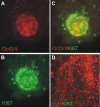
Figure 3. Some of the ES cell-derived neuronal colonies still contain at day 14 OCT3/4 and Ki67-positive cells.
(A) After 14 days of differentiation culture few colonies (less than 5%), as exemplified here, still contain OCT3/4-positive cell clusters as detected by immunofluorescence staining using a specific mAb (magnification 40×). (B) The same colony was stained by an anti-Ki67 mAb to detect proliferating cells. (C) The merged staining indicates that Ki67-positive proliferating cells exist which are not OCT3/4-positive stem cells. (D) In parallel, cells of the neuronal differentiation culture were stained at day 14 by anti-Ki67 and anti-Tuj1 mAb. The merged staining (magnification 20×) indicates that the vast majority of the cells are Tuj1-positive neuronal cells. Few Ki67-positive proliferating cells are negative for the neuronal cell marker.
Figure 4
Figure 4. Immunohistochemical staining identifies neuronal and glial cells in teratomas derived from ES and in vitro differentiated cells.
The tumors were grown in SCID/beige mice after injection of ES cells (A, C) or after injection of in vitro differentiated cells (B, D). The teratomas were stained by immunohistochemistry for the neuronal marker NeuN (A, B) and the glial marker GFAP (C, D) which is mainly found in astrocytes. In the sections shown here, groups of cells positive for NeuN and confluent groups of cells positive for GFAP were found.
Figure 5
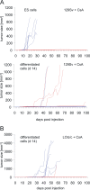
Figure 5. Tumor growth in CsA-treated 129Sv mice and LOU/c rats after injection of ES cells or in vitro differentiated cells.
(A) 1×10⁶ ES cells or in vitro differentiated cells were injected subcutaneously at day 0 into syngeneic 129Sv mice (for the numbers of animals see Table 2) which were treated daily with CsA (10 mg/kg body weight). The tumor size was recorded every second day until day 100 using linear calipers. The growth of tumors in individual mice is shown. Tumor growth in female hosts is indicated by red lines and male hosts by blue lines. (B) 1×10⁶ cells differentiated in vitro for 14 days were injected subcutaneously into LOU/c rats (n = 18) which were treated daily with CsA (10 mg/kg body weight). The tumor size was recorded every second day until day 100 using linear calipers. The growth of tumors in individual rats is shown. Tumor growth in female hosts is indicated by red and in male hosts by blue lines. One tumor in a female rat was not progressive and remained very small until the end of the experiment. In two male rats a tumor regression occurred.
Figure 6
Figure 6. Lysis of ES and in vitro differentiated cells by NK cells derived from naïve LOU/c rats and expression analysis of MHC class I molecules and NKG2D ligands.
(A) Mean of specific lysis and standard deviation (SD) of triplicates of ES or in vitro differentiated cells at three effector∶target (E∶T) ratios. Effector cells were lymphocytes obtained from spleens of 10 individual LOU/c rats by density gradient centrifugation on Biocoll. Results obtained with lymphocytes from female rats are indicated by open symbols and from male rats by filled symbols. (B) The mean specific lysis and SD of triplicates of ES, YAC-1, and RMA target cells at three effector∶target (E∶T) ratios by freshly isolated NK cells or NK cell-depleted splenocytes from naïve LOU/c rats are shown. The NK cell enriched fraction contained 86% and the NK cell depleted fraction 3% NKR-P1A-positive cells as determined by flow cytometry. The results are representative for 3 independent experiments. (C) The mean specific lysis and SD of triplicates of ES and YAC-1 cells at three effector∶target (E∶T) ratios by freshly isolated (open symbols) or 3 days in vitro with 1000 U/ml IL-2 stimulated (closed symbols) mouse and rat NK cells are shown. The NK cell-enriched fractions contained more than 80% NK cells as determined by flow cytometry. The results shown are representative for 3 independent experiments. (D) The expression of MHC class I molecules on ES and differentiated cells was analyzed by flow cytometry using anti-H2Kb and anti-H2Db Abs (full lines). The stainings with the isotype control are shown by the dotted lines. RMA cells (H2b) served as positive control for these antibodies. NKG2D ligands were stained with a recombinant mouse NKG2D-Fc chimeric protein and a FITC-conjugated goat anti-human IgG antibody as secondary reagent (full lines). Stainings with the secondary reagent only are shown by the dotted lines. YAC-1 cells (H2a) served as positive controls for these stainings. The results shown are representative for more than 3 independent experiments. (E) ES and as positive control YAC-1 cells were stained with the NKG2D-Fc chimeric protein and with mAbs specific for the NKG2D ligands H60, MULT-1, and RAE-1. The mean+SD of the proportion of positive cells and the mean fluorescence intensity+SD determined in 6 independent experiments is shown. (F) The mean inhibition+SD of specific lysis of ES and YAC-1 cells by soluble mouse NKG2D is shown as determined in 3 experiments. The mean of specific lysis of the target cells by rat NK cells at an effector to target ratio of 20∶1 was determined as described above and adjusted to 100%. For inhibition of lysis a soluble mouse NKG2D protein was added to the test at a concentration of 3 µg/ml. The relative lysis of the target cells exposed to NKG2D was calculated. (G) The mean inhibition+SD of specific lysis of ES cells by soluble mouse NKG2D and various inhibitory antibodies against NKG2D ligands is shown. The mean of specific lysis of the target cells by IL-2 activated mouse NK cells at an effector to target ratio of 10∶1 was determined as described above and adjusted to 100%. For inhibition of lysis a soluble mouse NKG2D protein or the indicated mAbs were added to the test at a concentration of 3 µg/ml. The relative lysis of the target cells exposed to NKG2D or the mAbs was calculated.
Figure 7
Figure 7. Lysis of ES and in vitro differentiated cells by splenocytes derived from rats 6 weeks after intracerebral grafting of in vitro differentiated cells.
Mean of specific lysis and SD of triplicates of ES cells (A, C, E, G) or in vitro differentiated cells (B, D, F) at three effector∶target (E∶T) ratios. Effector cells were lymphocytes obtained by density gradient centrifugation on Biocoll from spleens of grafted (no. 113–117) or naïve (co1 and co2) DA rats (A, B), grafted (no. 118–122) or naïve (co1 and co2) LEW.1N rats (C, D), and grafted Wistar rats (no. 38, 39, 48, 49, 51, 52, 53, 54, 56, 57). The individual rats are indicated by symbols. (G) Mean of specific lysis and SD of triplicates of ES cells by lymphokine-activated killer (LAK) cells derived from splenocytes of Wistar rats (no. 38, 48, 49, 51, 52, 54, 56) after culture for 4 days in the presence of 100 U/ml IL-2.
Figure 8
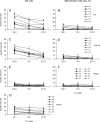
Figure 8. Analysis of effects of NK cells on the rejection of ES cells in vivo.
(A) 1×10⁶ ES cells were injected subcutaneously at day 0 into 15 T and B cell deficient SCID mice which have functional NK cells. The tumor size was recorded every second day until day 100 using linear calipers. The growth of tumors in individual mice is shown. Tumor growth in female hosts is indicated by red lines and in male hosts by blue lines. In one animal a tumor regression was observed. (B) The treatment scheme of rats receiving an NK cells depleting antibody (anti-NKR-P1A) or an isotype control is shown. (C) The mean proportion plus SD of NK cells in the blood of male LOU rats receiving the anti-NKR-P1A mAb (n = 8) or the isotype control (n = 6) is shown. Blood cells were stained with the respective mAb and analyzed by flow cytometry after lysis of erythrocytes. (D) The proportion of the male rats in which tumors were found at autopsy is indicated. The size of the tumors was 12 mm3 and 16 mm3, respectively. Both were palpable for more than 30 days before autopsy.

References

    1. Björklund LM, Sanchez-Pernaute R, Chung S, Andersson T, Chen IY, et al. Embryonic stem cells develop into functional dopaminergic neurons after transplantation in a Parkinson rat model. Proc Natl Acad Sci U S A. 2002;99:2344–2349. - PMC - PubMed
    1. Kim JH, Auerbach JM, Rodriguez-Gomez JA, Velasco I, Gavin D, et al. Dopamine neurons derived from embryonic stem cells function in an animal model of Parkinson's disease. Nature. 2002;418:50–56. - PubMed
    1. Baier PC, Schindehütte J, Thinyane K, Flugge G, Fuchs E, et al. Behavioral changes in unilaterally 6-hydroxy-dopamine lesioned rats after transplantation of differentiated mouse embryonic stem cells without morphological integration. Stem Cells. 2004;22:396–404. - PubMed
    1. Thinyane K, Baier PC, Schindehütte J, Mansouri A, Paulus W, et al. Fate of pre-differentiated mouse embryonic stem cells transplanted in unilaterally 6-hydroxydopamine lesioned rats: histological characterization of the grafted cells. Brain Res. 2005;1045:80–87. - PubMed
    1. Erdö F, Buhrle C, Blunk J, Hoehn M, Xia Y, et al. Host-dependent tumorigenesis of embryonic stem cell transplantation in experimental stroke. J Cereb Blood Flow Metab. 2003;23:780–785. - PubMed

Publication types