

Biatrial myxoma and cerebral ischemia successfully treated with intravenous thrombolytic therapy and surgical resection
- PMID: 18612442
- PMCID: PMC2435448
Biatrial myxoma and cerebral ischemia successfully treated with intravenous thrombolytic therapy and surgical resection
Abstract
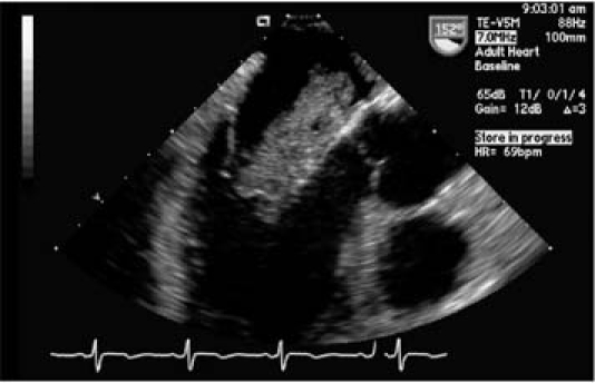
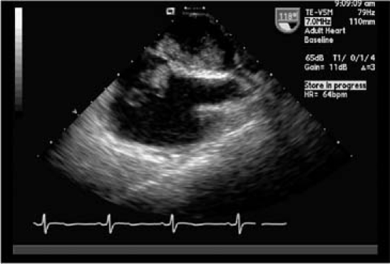
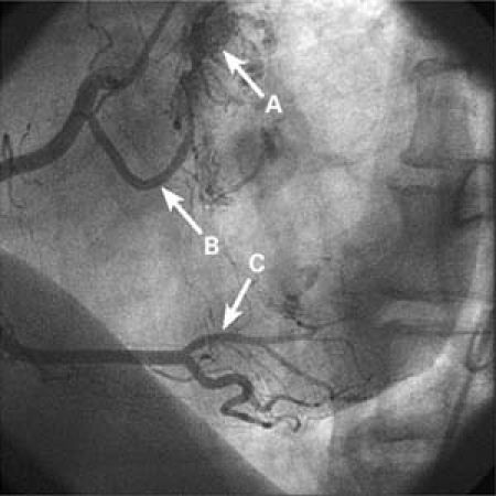
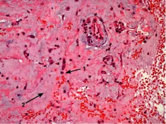
We report what we believe is the 1st case in the medical literature in which an intravenous thrombolytic agent was used successfully--without massive intracranial bleeding--to treat acute stroke induced by atrial myxoma. Our patient, who had biatrial myxomas with a dual blood supply from the right coronary artery, presented with cerebral ischemia. Transesophageal echocardiography was essential in clarifying the diagnosis and in helping to direct surgical treatment.
Keywords: Cerebrovascular accident; echocardiography, transesophageal; embolism; heart atria/surgery; heart neoplasms/complications/diagnosis/surgery; intracranial embolism and thrombosis/etiology/drug therapy; myxoma; thrombolytic therapy; tissue plasminogen activator.
Figures
References
-
- Molina JE, Edwards JE, Ward HB. Primary cardiac tumors: experience at the University of Minnesota. Thorac Cardiovasc Surg 1990;38 Suppl 2:183–91. - PubMed
-
- Tazelaar HD, Locke TJ, McGregor CG. Pathology of surgically excised primary cardiac tumors. Mayo Clin Proc 1992; 67(10):957–65. - PubMed
-
- Larrieu AJ, Jamieson WR, Tyers GF, Burr LH, Munro AI, Miyagishima RT, et al. Primary cardiac tumors: experience with 25 cases. J Thorac Cardiovasc Surg 1982;83(3):339–48. - PubMed
-
- Odim J, Reehal V, Laks H, Mehta U, Fishbein MC. Surgical pathology of cardiac tumors. Two decades at an urban institution. Cardiovasc Pathol 2003;12(5):267–70. - PubMed
-
- Kamiya H, Yasuda T, Nagamine H, Sakakibara N, Nishida S, Kawasuji M, Watanabe G. Surgical treatment of primary cardiac tumors: 28 years' experience in Kanazawa University Hospital. Jpn Circ J 2001;65(4):315–9. - PubMed
Publication types
MeSH terms
Substances
LinkOut - more resources
Full Text Sources