. 2008 Sep;316(1-2):177-86.
doi: 10.1007/s11010-008-9831-4. Epub 2008 Jul 9.

Inhibition of protein kinase CK2 suppresses angiogenesis and hematopoietic stem cell recruitment to retinal neovascularization sites

Affiliations

Inhibition of protein kinase CK2 suppresses angiogenesis and hematopoietic stem cell recruitment to retinal neovascularization sites

A A Kramerov et al. Mol Cell Biochem. 2008 Sep.

Abstract

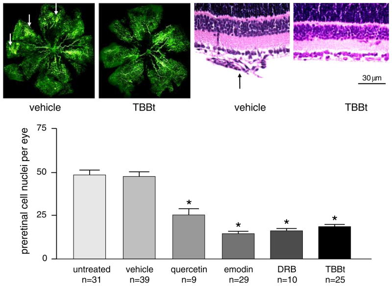
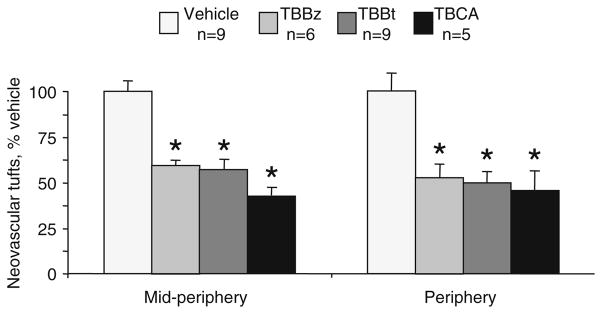
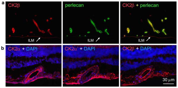
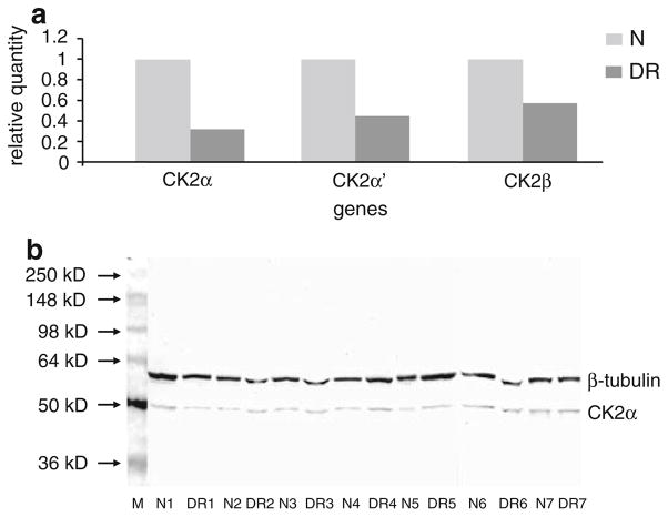
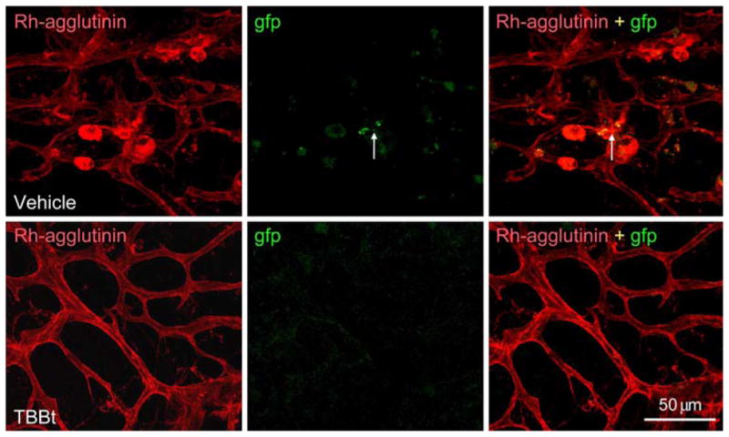
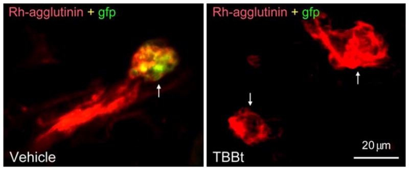
Ubiquitous protein kinase CK2 participates in a variety of key cellular functions. We have explored CK2 involvement in angiogenesis. As shown previously, CK2 inhibition reduced endothelial cell proliferation, survival and migration, tube formation, and secondary sprouting on Matrigel. Intraperitoneally administered CK2 inhibitors significantly reduced preretinal neovascularization in a mouse model of proliferative retinopathy. In this model, CK2 inhibitors had an additive effect with somatostatin analog, octreotide, resulting in marked dose reduction for the drug to achieve the same effect. CK2 inhibitors may thus emerge as potent future drugs aimed at inhibiting pathological angiogenesis. Immunostaining of the retina revealed predominant CK2 expression in astrocytes. In human diabetic retinas, mRNA levels of all CK2 subunits decreased, consistent with increased apoptosis. Importantly, a specific CK2 inhibitor prevented recruitment of bone marrow-derived hematopoietic stem cells to areas of retinal neovascularization. This may provide a novel mechanism of action of CK2 inhibitors on newly forming vessels.


Figures

Fig. 1
Fig. 1
Effect of CK2 inhibitors on growth factor-induced BREC migration. Confluent BREC were wounded and cultured for 7 days in 0.5% serum-containing medium with IGF-I + FGF-2 + VEGF + PlGF (four GFs) at 10 ng/ml each ± CK2 inhibitors, emodin (10 μM) and DRB (15 μM). Cell migration into the wound was counted using the AAB software. Error bars correspond to mean ± standard error of mean of at least three individual experiments. * P < 0.005 of CK2 inhibitor versus four GFs. Both inhibitors significantly reduced cell migration. Modified from [15]
Fig. 2
Fig. 2
CK2 inhibitors reduce retinal neovascularization in the OIR mouse model. Upper left pair, fluorescein-dextran perfusion of retinas from mice treated intraperitoneally with solvent (vehicle) or inhibitor (TBBt). Note reduction of brightly fluorescent vessel tufts (arrows) after TBBt treatment; upper right pair, hematoxylin-eosin-stained sections of retinas from mice treated with vehicle or TBBt. Arrow, neovascular tuft in vehicle-treated retinas; after treatment, the retina appears normal. Bar = 30 μm; lower graph, counts of preretinal nuclei in various groups of mice. Neovascularization is significantly decreased in all inhibitor-treated groups. Quercetin was used at 10 or 100 mg/kg/day with similar results. Emodin and DRB were used at 30 or 60 mg/kg/day with somewhat stronger inhibition at a higher dose, and TBBt, at 60 mg/kg/day. N, number of animals in a group. Five to ten sections per eye from each mouse were counted. * P < 0.001 versus untreated or vehicle. Error bars correspond to mean ± standard error of mean of at least three individual experiments. Adapted with modifications from [15]
Fig. 3
Fig. 3
Effects of specific CK2 inhibitors on retinal neovascularization. Following the OIR model, retinal flatmounts were stained with endothelium-specific rhodamine-conjugated Ricinus communis agglutinin and neovascular tufts were counted. The counts in the inhibitor-treated retinas are shown as percentage of those in vehicle-treated retinas taken as 100%. All three inhibitors, TBBt, TBBz, and TBCA, significantly reduced the number of neovascular tufts at 100 mg/kg/day. N, number of eyes in a group. * P < 0.01 versus vehicle. Error bars correspond to mean ± standard deviation
Fig. 4
Fig. 4
Schematic of intracellular signaling from angiogenic growth factors, somatostatin, insulin, and ECM. Somatostatin (SST; octreotide is its analog) and CK2 modulate several major signaling pathways involving growth factor receptor tyrosine kinases, G-protein coupled receptors (SSTR), and extracellular matrix (ECM) receptors, integrins. Along with many similarities in the action of these major effectors, there are notable differences. CK2 modulates Raf-ERK-S6K, p38 MAPK, and Akt pathways, whereas, octreotide can influence p27, STAT3, and PKA signaling. Together, SST/octreotide and CK2 can influence most major signaling pathways. This makes them good candidates for combination therapy for abnormal retinal angiogenesis
Fig. 5
Fig. 5
Combination therapy for retinal neovascularization. (a) Octreotide reduced preretinal neovascularization by 67% at 5 mg/kg/day and by 50% at 1 mg/kg/day. Emodin at 30 mg/kg/day showed 57% inhibition. When 1 mg/kg/day octreotide was combined with 30 mg/kg/day emodin, the same inhibitory effect was observed as with 5 mg/kg/day octreotide (69%). Combination of 30 mg/kg/day TBBt with 1 mg/kg/day octreotide produced 61% inhibition compared to 46% by TBBt alone. Five mice were used per each group in three independent experiments. Counts of preretinal nuclei as a measure of neovascularization in various groups of mice are shown. Vehicle represents emodin solvent because octreotide solvent was just PBS. Error bars correspond to mean ± standard deviation. * P < 0.001 versus vehicle, ** P < 0.05 versus single drug or vehicle. Combination of emodin or TBBt with octreotide more efficiently reduces mouse retinal neovascularization than any single drug alone; (b) representative cryostat sections of control and treated retinas. Immunofluorescent staining was performed with anti-perlecan (green color) to reveal the blood vessels and inner limiting membrane (ILM; located at the boundary of the retina and vitreous body), with counterstaining of nuclei with DAPI (blue color). Prominent preretinal vessels in the vehicle group (arrowheads) disappear after the treatments but much less change is seen in intraretinal vasculature. ONL, outer nuclear layer; INL, inner nuclear layer; GCL, ganglion cell layer. Bar = 60 μm. Adapted with modifications from [18] with permission from the American Society for Investigative Pathology
Fig. 6
Fig. 6
Positive CK2 staining of retinal vessels after epitope unmasking. (a) Double staining of human retinal section for CK2β (red color) and perlecan (green color). There is co-distribution of both labels in retinal capillaries (right panel, yellow color). In this particular small area astrocytes that are also positive for CK2 were virtually absent. ILM, inner limiting membrane. (b) Staining of serial human retinal sections for different subunits of CK2. A large vessel is positive (red color) along with astrocytes in the inner retina. CK2α and β subunits give identical staining, but α′ subunit has less apical and more basal staining in the vessel wall. Nuclei were counterstained with DAPI (blue color)
Fig. 7
Fig. 7
Expression of CK2 subunits in normal and diabetic human retina. (a) Real-time RT-PCR of different CK2 subunits. Note decreased levels of all messages in the diabetic retinopathy (DR) group compared to age-matched non-diabetic group (N). β-micro-globulin was used as a normalizing housekeeping gene [18]. (b) Western blot of individual retinal samples including seven non-diabetic (N) and seven with diabetic retinopathy (DR). The membrane was probed with anti-CK2α antibody. Lane loading was normalized by β-tubulin. There are no significant differences in the amount of CK2α between diabetic and non-diabetic samples. Markers in kD are at the left
Fig. 8
Fig. 8
HSC incorporation into sites of retinal neovascularization in the mouse OIR model. (a and b) Mouse retinal flat mount double stained with rhodamine-agglutinin (Rh-agglutinin) to reveal blood vessels and anti-gfp to reveal gfp+ c-kit+, Sca-1+ HSC (arrowheads) injected intravitreally. Arrows point to neovascular tufts in the peripheral retina. Bar = 100 μm; (c) superimposition of pictures in a and b at higher magnification. Several neovascular tufts (arrows) contain gfp + HSC. These appear in yellow color. Bar = 50 μm
Fig. 9
Fig. 9
Effect of TBBt on HSC incorporation into mouse retinal neovasculature. Vehicle, double-stained retinal flat mount of vehicle-treated retina. Note several neovascular tufts revealed by rhodamine-agglutinin; two of them contain gfp+ HSC (arrow); TBBt, no tufts and no gfp+ HSC are visible after TBBt treatment (5 days at 100 mg/kg/day). Bar = 50 μm
Fig. 10
Fig. 10
Lack of HSC incorporation into neovasculature upon TBBt treatment. Vehicle, vehicle-treated retina at high magnification. A rhodamine-agglutinin positive (red color) neovascular tuft (arrow) contains several gfp+ HSC (green-yellow color); TBBt, two neovascular tufts (arrows) remaining after TBBt treatment are shown; they are both devoid of gfp+ HSC. Bar = 20 μm

References

    1. Meggio F, Pinna LA. One-thousand-and-one substrates of protein kinase CK2? FASEB J. 2003;17:349–368. - PubMed
    1. Unger GM, Davis AT, Slaton JW, et al. Protein kinase CK2 as regulator of cell survival: implications for cancer therapy. Curr Cancer Drug Targets. 2004;4:77–84. - PubMed
    1. Ahmad KA, Wang G, Slaton J, et al. Targeting CK2 for cancer therapy. Anticancer Drugs. 2005;16:1037–1043. - PubMed
    1. Sarno S, Salvi M, Battistutta R, Zanotti G, Pinna LA. Features and potentials of ATP-site directed CK2 inhibitors. Biochim Biophys Acta. 2005;1754:263–270. - PubMed
    1. Canton DA, Litchfield DW. The shape of things to come: an emerging role for protein kinase CK2 in the regulation of cell morphology and the cytoskeleton. Cell Signal. 2006;18:267–275. - PubMed

Publication types

MeSH terms