. 2008 Jul 8;6(7):e167.
doi: 10.1371/journal.pbio.0060167.

A short-time scale colloidal system reveals early bacterial adhesion dynamics

Affiliations

A short-time scale colloidal system reveals early bacterial adhesion dynamics

Christophe Beloin et al. PLoS Biol. .

Abstract

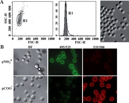
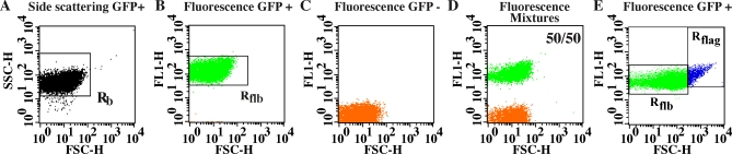
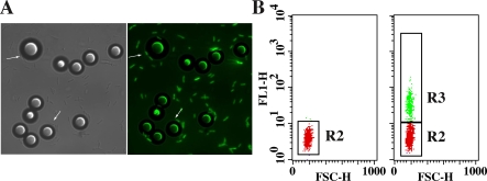
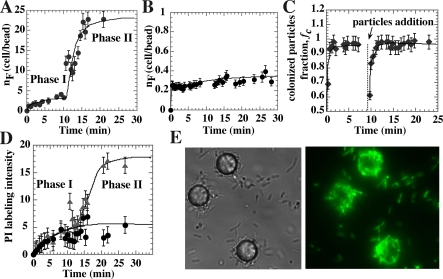
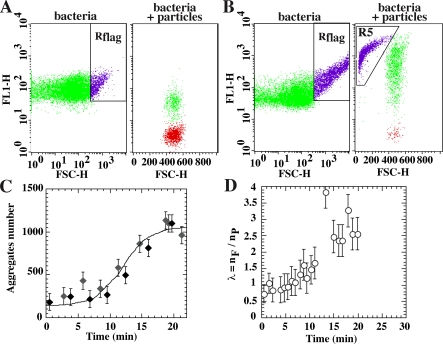
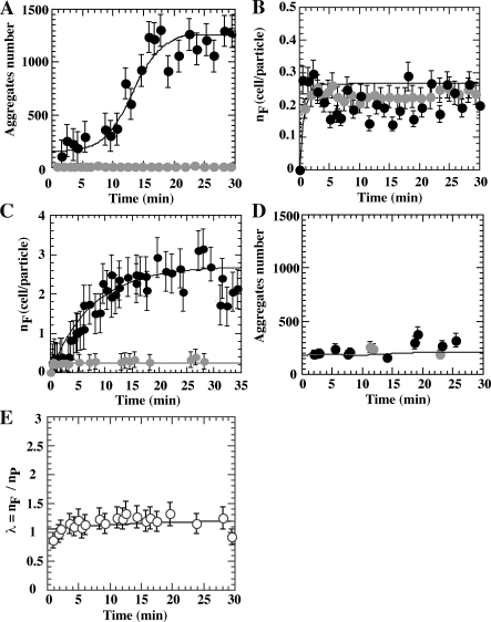
The development of bacteria on abiotic surfaces has important public health and sanitary consequences. However, despite several decades of study of bacterial adhesion to inert surfaces, the biophysical mechanisms governing this process remain poorly understood, due, in particular, to the lack of methodologies covering the appropriate time scale. Using micrometric colloidal surface particles and flow cytometry analysis, we developed a rapid multiparametric approach to studying early events in adhesion of the bacterium Escherichia coli. This approach simultaneously describes the kinetics and amplitude of early steps in adhesion, changes in physicochemical surface properties within the first few seconds of adhesion, and the self-association state of attached and free-floating cells. Examination of the role of three well-characterized E. coli surface adhesion factors upon attachment to colloidal surfaces--curli fimbriae, F-conjugative pilus, and Ag43 adhesin--showed clear-cut differences in the very initial phases of surface colonization for cell-bearing surface structures, all known to promote biofilm development. Our multiparametric analysis revealed a correlation in the adhesion phase with cell-to-cell aggregation properties and demonstrated that this phenomenon amplified surface colonization once initial cell-surface attachment was achieved. Monitoring of real-time physico-chemical particle surface properties showed that surface-active molecules of bacterial origin quickly modified surface properties, providing new insight into the intricate relations connecting abiotic surface physicochemical properties and bacterial adhesion. Hence, the biophysical analytical method described here provides a new and relevant approach to quantitatively and kinetically investigating bacterial adhesion and biofilm development.


Conflict of interest statement

Competing interests. The authors have declared that no competing interests exist.

Figures

Figure 1
Figure 1. Colloidal Substrates
(A) FCM scattering plot and corresponding bright field microscope images of a COO particle sample washed in water and suspended in PBS. 80% of events were concentrated in the scattering value region R1 corresponding to a monodispersed, 10-μm-diameter population. Outliers corresponded to a population of slightly larger particles and a few doublets. Cationic (NH3 +) particles displayed the same scattering characteristics as anionic (COO) particles. (B) Microscopic images of PYR- (2nd column) and PI- (3rd column) labeled cationic (1st row) and anionic (2nd row) particles. Dye concentration equal to 16.7 nM.
Figure 2
Figure 2. Bacterial FCM
Scattering (A) and fluorescence dot plots of all GFP-labeled (B), all non GFP-labeled cells (C) and a mixture (50/50) of both (D). Aliquots of exponentially growing cultures were diluted in PBS to obtain a 5 × 10⁶ cells/ml suspension. (E) FL1 dot plots of a slightly aggregated cell population (Rflag, right plot); in this case, 3% of the objects are considered aggregates.
Figure 3
Figure 3. Bacterial Adhesion in Colloidal Suspension
(A) Microscope images of MG1655gfp E. coli cells and particles brought into contact with particles in M63B1 medium for 20 min. Bright-field and bright-field fluorescence combined images are shown. Adhesive events can be observed on the combined image; examples are indicated by white arrows. (B) Corresponding fluorescence dot plots are shown for particles alone (left) and cells and particles brought into contact for 20 min (right). Gate R2 corresponds to bare particles, whereas gate R3 corresponds to colonized particles.
Figure 4
Figure 4. Effect of Curli Production on Early Surface Adhesion
Adhesion kinetic curves of MG1655gfpompR234 (A) and MG1655gfpΔcsgA (B). Particles and cells were brought into contact at time t = 0. Cell to particle ratio was around 200 and particle concentration equal to 6 × 10⁶/ml. (C) Colonized particle fraction kinetics are shown for two sequential additions of fresh NH3 + particles to MG1655gfpompR234 cells. 50 μl of stock particles were added to a cell/particle sample after 9 min incubation once the first colonization phase leveled off. (D) Charge inversion of NH3 + particle during surface colonization. FL3 intensity in time due to PI adsorption (2 × 10⁻⁸ M) is shown for MG1655gfpompR234 (▴) and MG1655gfpΔcsgA (•). (E) Bright field and fluorescence microscope images of MG1655gfpompR234 engaged in second-phase colonization of NH3 + particles.
Figure 5
Figure 5. Self-Association of Free-Floating Cells and Cooperative Effects in Curli-Producing Bacteria
Bacteria (left) and colloid (right) dot plots of MG1655gfpompR234 brought into contact with NH3 + particles before (A) and after (B) second colonization phase onset. (C) Number of events comprised in the Rflag gate in the presence (black diamond) or absence (gray diamond) of particles. Cells were taken from a culture (DO = 0.5) and resuspended by pipetting in an adequate volume of buffer at time t = 0. (D) Time dependence of cooperative index, λ, during MG1655gfpompR234 colonization.
Figure 6
Figure 6. Aggregation and Surface Colonization Kinetics of Cells Expressing Different Cell Surface Adhesins
Ag43 (A and B); F-pili (C and D) were examined under the same conditions as in Figure 4. (black circles) corresponds to cells producing indicated surface adhesin and (gray circles) to cells not producing surface adhesin. (E) Cooperative index in time for F pili–expressing cell colonization of NH3 + particles.

Comment in

References

    1. Hall-Stoodley L, Costerton JW, Stoodley P. Bacterial biofilms: from the natural environment to infectious diseases. Nat Rev Microbiol. 2004;2:95–108. - PubMed
    1. Donlan RM. Biofilms: microbial life on surfaces. Emerg Infect Dis. 2002;8:881–890. - PMC - PubMed
    1. Katsikogianni M, Missirlis YF. Concise review of mechanisms of bacterial adhesion to biomaterials and of techniques used in estimating bacteria-material interactions. Eur Cell Mater. 2004;8:37–57. - PubMed
    1. Bos R, van der Mei HC, Busscher HJ. Physico-chemistry of initial microbial adhesive interactions–its mechanisms and methods for study. FEMS Microbiol Rev. 1999;23:179–230. - PubMed
    1. de Kerchove AJ, Elimelech M. Relevance of electrokinetic theory for “soft” particles to bacterial cells: implications for bacterial adhesion. Langmuir. 2005;21:6462–6472. - PubMed

Publication types

MeSH terms