. 2008 Jul 11;134(1):37-47.
doi: 10.1016/j.cell.2008.05.049.

Highly efficient, functional engraftment of skeletal muscle stem cells in dystrophic muscles

Affiliations

Highly efficient, functional engraftment of skeletal muscle stem cells in dystrophic muscles

Massimiliano Cerletti et al. Cell. .

Abstract

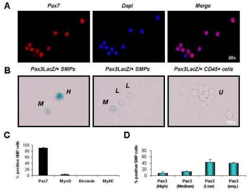
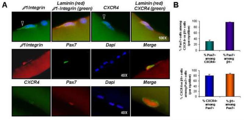
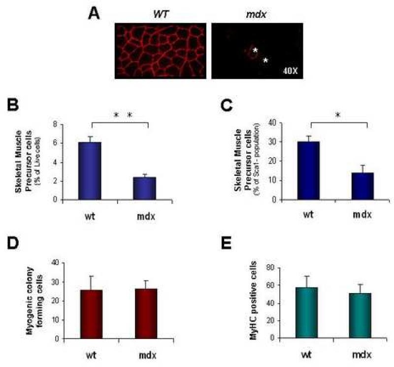
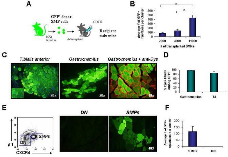
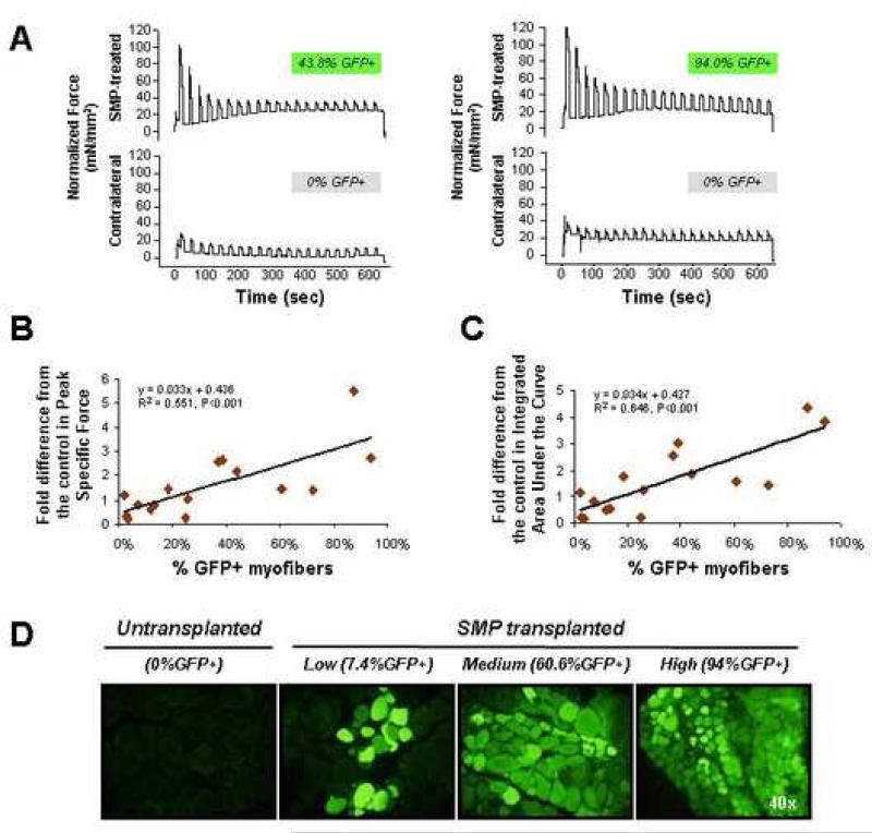
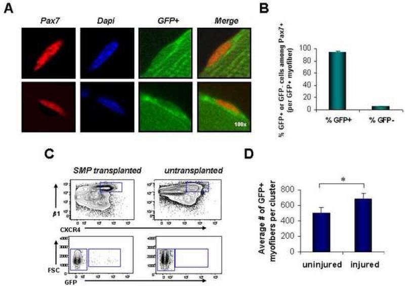
Satellite cells reside beneath the basal lamina of skeletal muscle fibers and include cells that act as precursors for muscle growth and repair. Although they share a common anatomical localization and typically are considered a homogeneous population, satellite cells actually exhibit substantial heterogeneity. We used cell-surface marker expression to purify from the satellite cell pool a distinct population of skeletal muscle precursors (SMPs) that function as muscle stem cells. When engrafted into muscle of dystrophin-deficient mdx mice, purified SMPs contributed to up to 94% of myofibers, restoring dystrophin expression and significantly improving muscle histology and contractile function. Transplanted SMPs also entered the satellite cell compartment, renewing the endogenous stem cell pool and participating in subsequent rounds of injury repair. Together, these studies indicate the presence in adult skeletal muscle of prospectively isolatable muscle-forming stem cells and directly demonstrate the efficacy of myogenic stem cell transplant for treating muscle degenerative disease.


Figures

FIGURE 1
FIGURE 1. SMPs express markers of undifferentiated satellite cells
(A) Double-sorted SMPs (see Figures S1 and S4) were stained with antibody against Pax7. 90% (± 3.2%) of freshly sorted SMP cells showed nuclear Pax7 protein (red). Cell nuclei marked by DAPI (blue). (B) SMPs or CD45+ hematopoietic cells (as a negative control) were sorted from pooled limb muscles (including EDL, TA, gastrocnemius, quadriceps, soleus, and triceps brachii) of adult Pax3LacZ/+ heterozygous reporter mice, which express one copy of the bacterial β-galactosidase gene from the endogenous Pax3 promoter, providing a reliable reporter for Pax3 expression (Relaix et al., 2003). Double-sorted cells were stained with X-gal to detect LacZ activity and scored as high expressers (H, see left panel), medium expressers (M, see left and middle panels), low expressers (L, see middle panel), or undetectable (U, see right panels; cells were scored as undetectable if the level of X-gal staining was equivalent to background staining of the control CD45+ cell population, which does not express Pax3 (data not shown)). (C) Freshly sorted SMPs were stained as in (A) with antibody against Pax7, MyoD, Desmin or MyHC. Most SMPs are Pax7+, and <5% were MyoD+. No expression of Desmin or MyHC was detected. (D) Quantification of Pax3 expression by sorted SMPs revealed heterogeneous levels of Pax3 expression, as expected based on previous studies (Montarras et al., 2005; Relaix et al., 2006). Data are presented as the frequency (Mean ± SD) of SMP cells exhibiting high, medium, low, or undetectable (neg.) LacZ activity.
FIGURE 2
FIGURE 2. Expression of β1-integrin and CXCR4 by skeletal muscle satellite cells
(A) Single myofibers isolated from gastrocnemius or soleus muscles of C57BL mice were stained with anti-β1-integrin (green, top row, and red, middle row) or anti-CXCR4 (green, top and bottom rows) antibody, together with anti-laminin (red, top row) or anti-Pax7 (green, middle row and red, bottom row) and analyzed by fluorescence microscopy. Nuclei were marked by DAPI (blue). Open arrowheads denote β1-integrin+ or CXCR4+ cells (top row). Enlarged, merged images are shown at right (middle and bottom rows). (B) The average frequency of Pax7 and CXCR4 or Pax7 and β1-integrin co-expression by myofiber-associated cells was determined from analysis of 60 individual myofibers. Bar graphs depict the frequency of CXCR4-expressing (green) or β1-integrin-expressing (purple) myofiber-associated cells that also express Pax7 (Mean ± SEM), or the frequency of Pax7+ cells that also express CXCR4 (blue) or β1-integrin (orange). The majority (80-90%) of Pax7+ cells are also CXCR4+ and β1-integrin+ (bottom plot). Likewise, >90% of β1-integrin+ cells are also Pax7+, although only ~30% of CXCR4+ cells co-express Pax7 (top plot). Based on flow cytometric analysis, CXCR4+ cells that lack Pax7 expression appear for the most part to be CD45+ infiltrating inflammatory cells (data not shown), which exhibit no myogenic activity.
FIGURE 3
FIGURE 3. SMP frequency is reduced in dystrophic mice
Dystrophic disease in mdx animals is characterized by acute degeneration and regeneration of skeletal muscle during the first month of life, followed by a mild chronic dystrophy that begins at ~6-8 wks. of age and lasts throughout life. (A) Immunostaining for dystrophin protein in wild-type (wt) C57BL/Ka mice reveals protein expression at the cell membrane of every myofiber. In contrast, the majority of myofibers in mdx mice lack dystrophin expression, with the exception of a small number (~1-6% of total myofibers in mice aged 10-20 wks (Wernig et al., 2005)) of revertant fibers generated by skipping of the mutated exon (Lu et al., 2000). Two revertant fibers in the mdx muscle section shown are marked by asterisks (*). (B and C) Flow cytometric analysis of SMP frequency in dystrophic mdx mice shows an ~3-fold decrease in the frequency of SMPs among myofiber-associated cells (B) and an ~2-fold decrease among Sca-1- myofiber-associated cells (C) harvested from mdx muscle. *P<0.05, **P<0.01 (D and E) Double-sorted SMPs from mdx mice exhibited equivalent in vitro clonal plating efficiency (D, frequency of colony formation from single cells, as in Figure S3) and differentiation capacity (E, % of MyHC-expressing cells after culture in differentiation medium, as in Figure S3) as compared to SMPs sorted from wild type (wt) mice.
FIGURE 4
FIGURE 4. SMPs robustly engraft skeletal muscle in vivo
(A) Experimental design. Double-sorted GFP+ SMPs were injected intramuscularly into recipient mdx mice injured 1 day previously by injection of cardiotoxin (CDTX) into the same muscle. (B) Quantitative analysis of donor-derived (GFP+) myofibers in TA muscles injected with 2,000 (n=3), 4,000 (n=3) or 11,000 (n=3) SMPs. Recipient muscles were harvested 4 wks. after transplantation and analyzed for GFP expression by direct epifluorescence microscopy of transverse muscle sections. The total number of GFP+ myofibers per section was determined for 100-300 sections taken throughout the muscle, in order to determine the maximal number of donor-derived fibers generated in each muscle. Data are plotted as the mean (± SEM) number of GFP+ myofibers detected in the section of each engrafted muscle that contained the most GFP+ myofibers. *P < 0.01. (C) Transverse frozen sections of TA (left panel) and gastrocnemius (middle and right panels) muscles obtained from mdx mice transplanted 4 wks. previously with 11,000 GFP+ SMPs showed large clusters of regenerating donor-derived myofibers (GFP+, shown in green) with characteristic centrally localized nuclei and restored dystrophin expression (shown in red; dystrophin staining is shown on the right image only). GFP detection by epifluorescence (as in C) was confirmed by indirect immunofluorescence and immunohistochemistry using anti-GFP antibodies (see Figure S5). (D) Quantification of the frequency (mean ± SD) of dystrophin+ myofibers among GFP+ donor-derived myofibers in the TA or gastrocnemius of mdx mice transplanted with 11,000 SMP cells per muscle, revealed that the majority (85-100%) of GFP+ myofibers co-stained with dystrophin protein (red), which normally is lacking on most mdx myofibers ((Sicinski et al., 1989) and see Figure 3). (E and F) Myofiber-associated cells lacking SMP markers do not generate myofibers when transplanted in vivo. CD45-Sca-1-Mac-1-CXCR4-β1-integrin- (double negative, DN) cells or CD45-Sca-1-Mac-1-CXCR4+β1-integrin+ SMPs were twice-sorted (to ensure purity) from β-actin/GFP mice and then transferred at equal cell number (4000 per muscle) into separate pre-injured mdx recipients. Four weeks after transplant, injected muscles were harvested and sectioned. No GFP+ myofibers were found in muscles transplanted with DN cells (n=3), while muscle receiving GFP+ SMPs showed efficient contribution of GFP+ myofibers (n=3).
FIGURE 5
FIGURE 5. Functional improvement in muscle contractile force following engraftment of wild-type SMPs into dystrophic muscle
(A) Representative traces from contractile force measurements of SMP-transplanted (top rows) or mock-transplanted muscles. Traces are paired showing force production from contralateral muscles of the same recipient animal, and are normalized for muscle cross-sectional area. The % GFP+ myofibers detected upon subsequent sectioning of each muscle is indicated above each trace. Normalized data for all animals in the study, used for calculation of the fold differences and regression curves shown in (B and C) are provided in Table S1. (B) Regression analysis shows a significant (P<0.001) correlation between the fold difference from the control in Peak Specific Force production for the 10 min contraction protocol (plotted as the ratio of the average peak specific force in SMP-transplanted vs. mock-transplanted, contralateral soleus muscle) and the % GFP+ myofibers in the transplanted muscle. Each diamond represents data from an individual mouse (n=16). (C) Regression analysis shows significant (P<0.001) correlation between the fold difference from the control in Integrated Area Under the Curve (amplitude X duration) for the 10 min contraction protocol (plotted as the average integrated area under the curve in SMP-transplanted vs. mock-transplanted, contralateral soleus muscle) and the % GFP+ myofibers in the engrafted muscle. Each diamond represents an individual mouse (n=16). (D) Representative epifluorescence images showing GFP expression (green) in myofibers of mock-transplanted (left-most panel) or SMP-transplanted (three right panels) soleus muscles of individual mdx mice. Individual soleus muscles showed highly variable levels of engraftment, likely related to technical limitations in the efficiency of cell delivery that arise from the small size and less accessible anatomical location of the soleus.
FIGURE 6
FIGURE 6. Transplanted SMPs re-seed the satellite cell compartment
(A) Intact myofibers isolated from the gastrocnemius and soleus muscles of previously transplanted mdx mice show donor-derived GFP+ Pax7+ satellite cells associated with GFP+ myofibers. GFP is shown in green, Pax7 in red, and DAPI-stained nuclei in blue. Two representative image series are shown. (B) Quantitative analysis of the frequency of GFP+ and GFPPax7+ cells detected on single GFP+ myofibers isolated from previously transplanted mdx muscles (gastrocnemius and soleus, n=41 individual myofibers). The majority of Pax7+ cells on GFP+ myofibers are GFP+. (C) Re-isolation of GFP+ SMPs from the myofiber-associated cell compartment of mdx mice transplanted 4 wks. previously with GFP+ SMPs. The top contour plots depict events already gated for forward and side-scatter, live (calcein+ propidium iodide-), Sca-1-CD45-Mac-1- cells, and indicates the CXCR4+β1-integrin+ SMP population, which is further analyzed for GFP expression in the lower plot. <1% of re-isolated SMPs were GFP+, in part due to low total numbers of GFP+ myofibers in this particular cohort of mice (data not shown, n=4). GFP+ SMPs were not detected in untransplanted control muscles (right panels). (D) TA muscles from mdx mice previously transplanted with GFP+ SMPs were injured by cardiotoxin injection 4 weeks after transplantation. Analysis of donor-derived myofibers was performed 1 week after injury by direct epifluorescence microscopy. Data are presented as the mean (± SD) number of GFP+ myofibers detected in the section of each engrafted muscle that contained the most GFP+ myofibers (as in Figure 4). Re-injured muscles showed a significantly greater number of donor-derived myofibers (n=3), as compared to uninjured control muscles that were not re-injured (n=3). *P < 0.05.

Comment in

References

    1. Bachrach E, Perez AL, Choi YH, Illigens BM, Jun SJ, del Nido P, McGowan FX, Li S, Flint A, Chamberlain J, Kunkel LM. Muscle engraftment of myogenic progenitor cells following intraarterial transplantation. Muscle Nerve. 2006;34:44–52. - PubMed
    1. Beauchamp JR, Heslop L, Yu DS, Tajbakhsh S, Kelly RG, Wernig A, Buckingham ME, Partridge TA, Zammit PS. Expression of CD34 and Myf5 defines the majority of quiescent adult skeletal muscle satellite cells. J Cell Biol. 2000;151:1221–1234. - PMC - PubMed
    1. Blau HM, Webster C, Pavlath GK. Defective myoblasts identified in Duchenne muscular dystrophy. Proc Natl Acad Sci U S A. 1983;80:4856–4860. - PMC - PubMed
    1. Buckingham M. Myogenic progenitor cells and skeletal myogenesis in vertebrates. Curr Opin Genet Dev. 2006;16:525–532. - PubMed
    1. Collins CA, Olsen I, Zammit PS, Heslop L, Petrie A, Partridge TA, Morgan JE. Stem cell function, self-renewal, and behavioral heterogeneity of cells from the adult muscle satellite cell niche. Cell. 2005;122:1–13. - PubMed

Publication types