. 2008 Jul 11;134(1):175-87.
doi: 10.1016/j.cell.2008.05.045.

Distinct role of long 3' UTR BDNF mRNA in spine morphology and synaptic plasticity in hippocampal neurons

Affiliations

Distinct role of long 3' UTR BDNF mRNA in spine morphology and synaptic plasticity in hippocampal neurons

Juan Ji An et al. Cell. .

Abstract

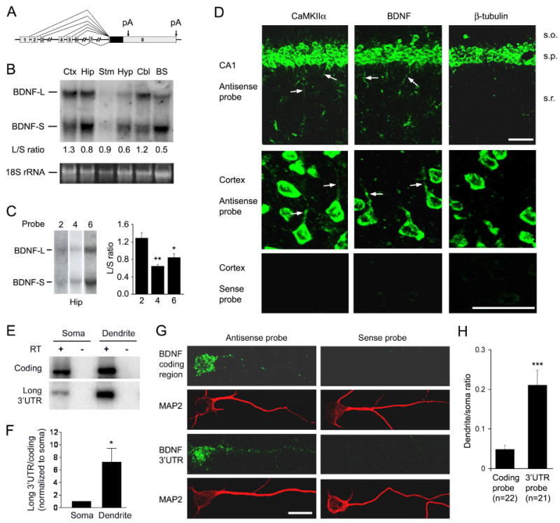
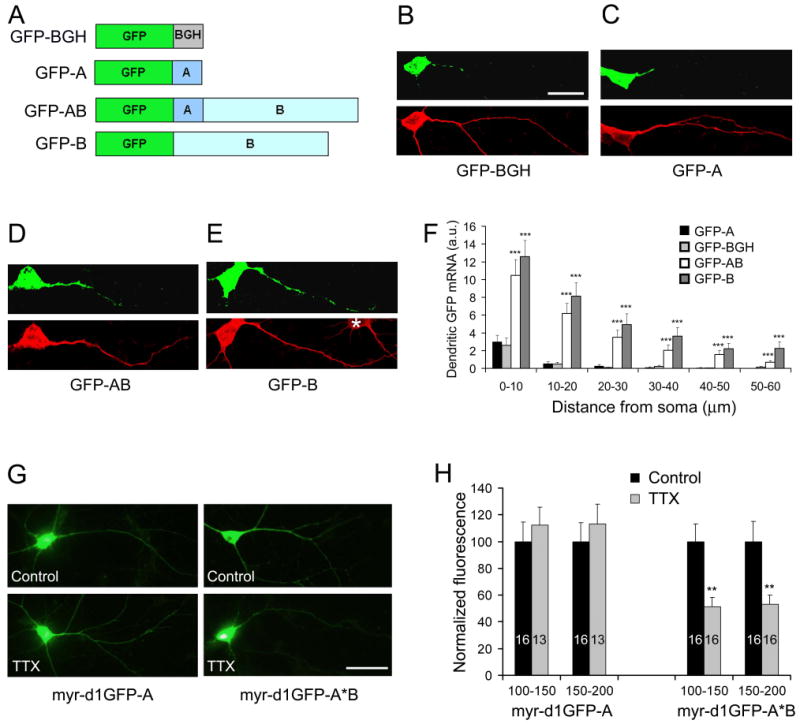
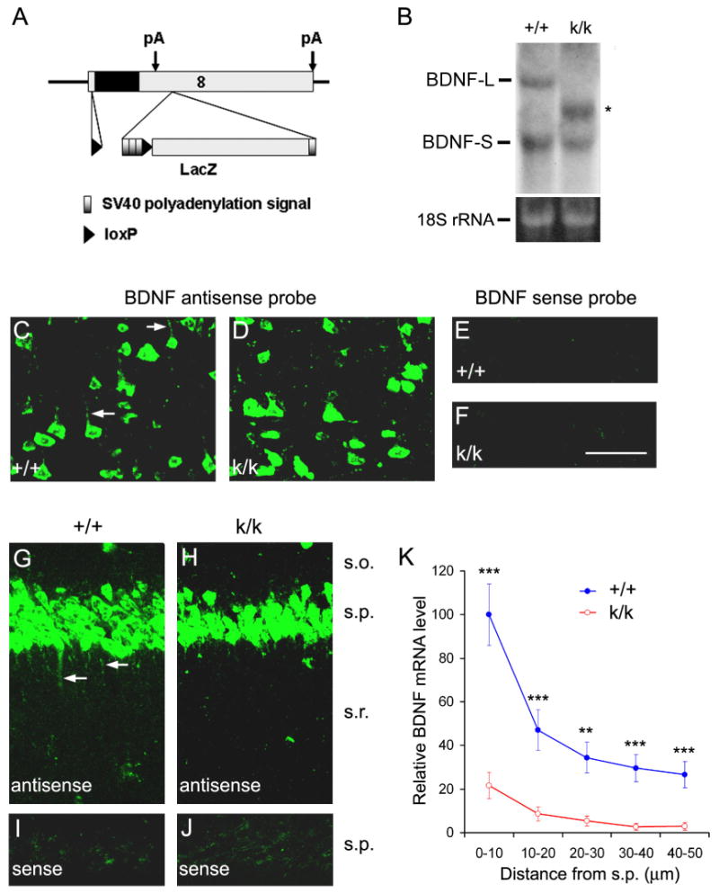
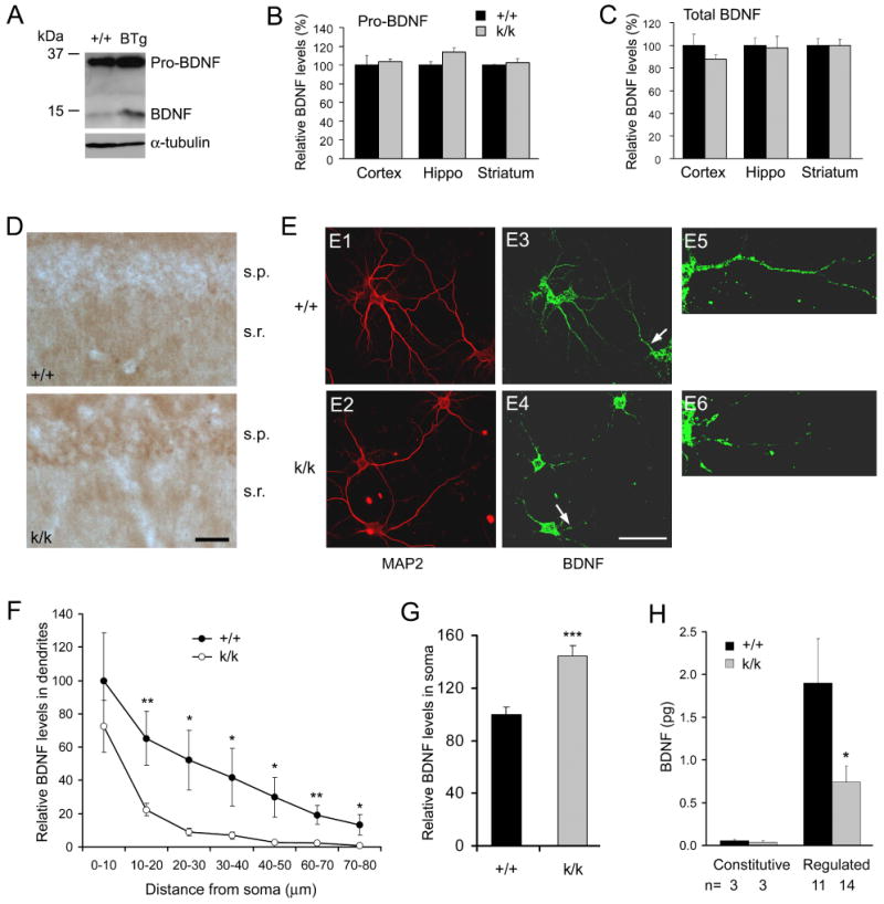
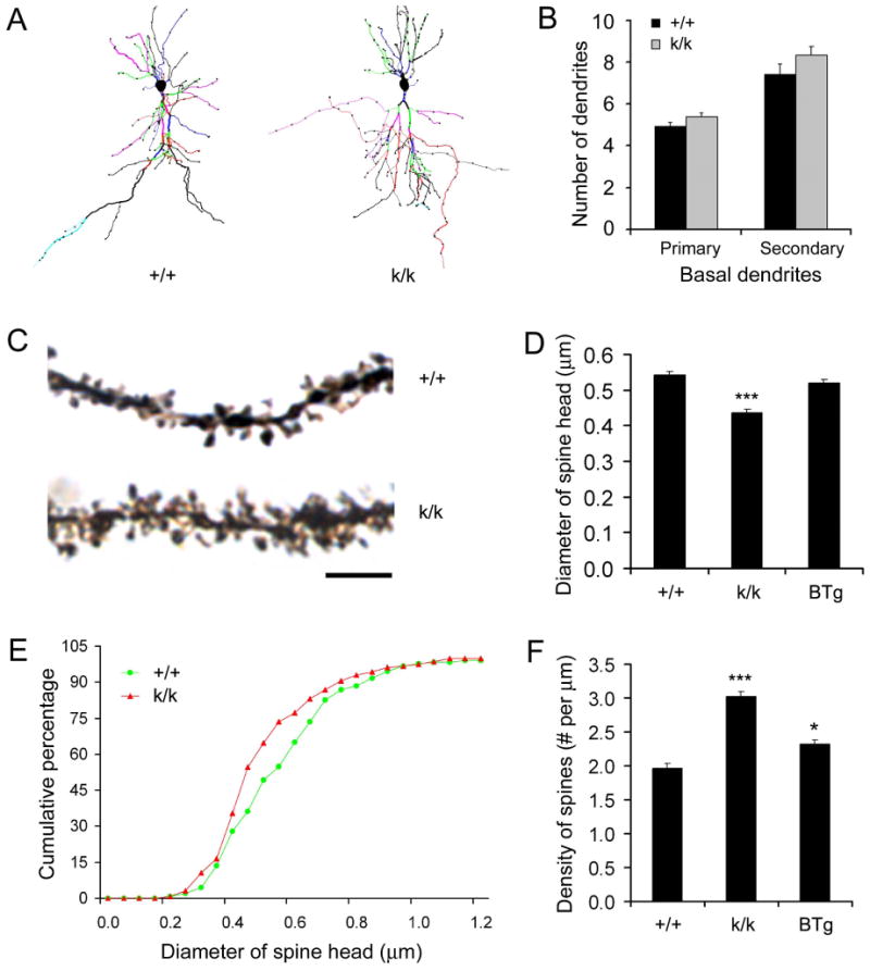
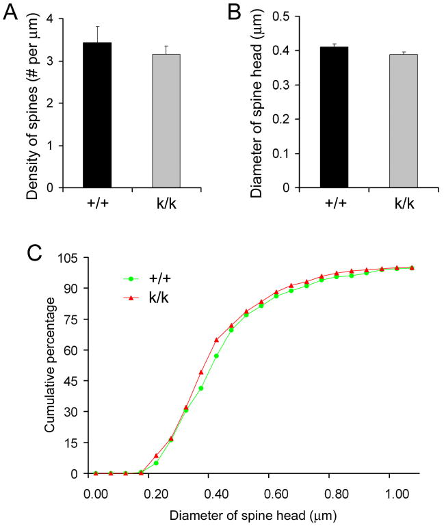
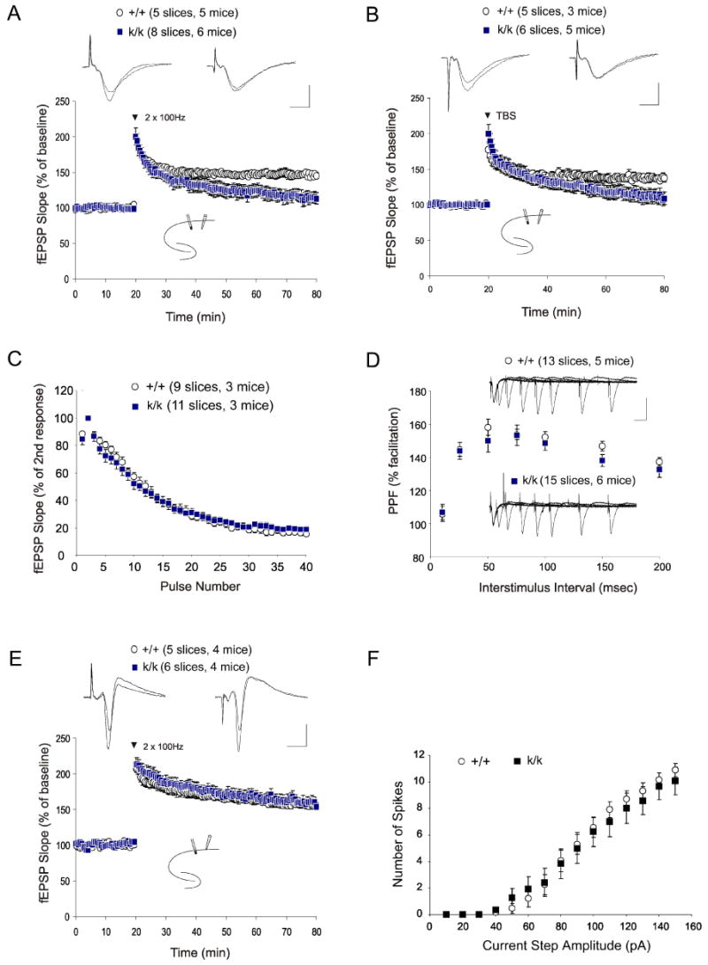
The brain produces two brain-derived neurotrophic factor (BDNF) transcripts, with either short or long 3' untranslated regions (3' UTRs). The physiological significance of the two forms of mRNAs encoding the same protein is unknown. Here, we show that the short and long 3' UTR BDNF mRNAs are involved in different cellular functions. The short 3' UTR mRNAs are restricted to somata, whereas the long 3' UTR mRNAs are also localized in dendrites. In a mouse mutant where the long 3' UTR is truncated, dendritic targeting of BDNF mRNAs is impaired. There is little BDNF in hippocampal dendrites despite normal levels of total BDNF protein. This mutant exhibits deficits in pruning and enlargement of dendritic spines, as well as selective impairment in long-term potentiation in dendrites, but not somata, of hippocampal neurons. These results provide insights into local and dendritic actions of BDNF and reveal a mechanism for differential regulation of subcellular functions of proteins.


Figures

Figure 1
Figure 1. Localization of long 3′UTR BDNF mRNA to dendrites
(A) Diagram of the mouse Bdnf gene depicting two alternative polyadenylation (pA) sites in exon 8 (arrows). Curved lines linking boxes (exons) indicate alternative splicing from the first seven exons to exon 8. The filled box within exon 8 represents the coding sequence. (B) Long and short 3′UTR BDNF mRNAs in different brain regions. BDNF mRNAs with a short 3′UTR (BDNF-S) or a long 3′UTR (BDNF-L) were present in the cortex (Ctx), hippocampus (Hip), striatum (Stm), hypothalamus (Hyp), cerebellum (Cbl), and brainstem (BS). Lower panel: 18S rRNA as a loading control. (C) Ratios of long and short BDNF mRNAs (L/S) derived from different Bdnf promoters. The same blot was used for all probes (n=4 mice). (D) Localization of BDNF mRNAs in dendrites of hippocampal CA1 and cortical neurons. Riboprobes were derived from the coding regions for CaMKIIα, BDNF, and β-tubulin. CaMKIIα and BDNF mRNAs were detected in dendrites (arrows). Abbreviations in this and all other figures: s.o., stratum oriens; s.p., stratum pyramidale; s.r., stratum radiatum. Scale bars, 50 μm. (E) Relative abundance of the long 3′UTR BDNF mRNA in the soma and dendrite fractions. (F) Ratio of the long 3′UTR signal to the coding region signal in the soma and dendrite fractions (n=3). (G) Distribution of BDNF mRNA in cell bodies and dendrites of cultured rat cortical neurons. MAP2 immunostaining marks cell bodies and dendrites. Scale bar, 20 μm. (H) Ratio of FISH signals in dendrites to those in cell bodies.
Figure 2
Figure 2. Cis-acting sequence in the 3′UTR required for the dendritic localization
(A) Schematic of four GFP constructs with different 3′UTRs. (B-E) Localization of GFP mRNA in neurons transfected with GFP-BGH (B), GFP-A (C), GFP-AB (D), or GFP-B (E). Top panels: FISH of cultured neurons with a GFP antisense riboprobe; Bottom panels: MAP2 immunocytochemistry; *, an untransfected neuron; Scale bar, 25 μm. (F) Levels of GFP mRNAs along the main apical dendrites of transfected neurons. Data were obtained from 15-23 transfected neurons grown on 3 coverslips for each construct and presented in arbitrary unit (a.u.). GFP-A was compared with the other three constructs. (G) Whole cell images of cultured hippocampal neurons expressing either myr-d1GFP-A or myr-d1GFP-A*B. The images were taken with a fluorescent microscope using a GFP filter. Scale bar, 50 μm. (H) Quantification of myr-d1GFP fluorescence in dendrites. Fluorescence intensities on distal dendrites (100-150 μm and 150-200 μm away from somata) were measured and normalized to control levels. The number of neurons for each condition is indicated inside bars.
Figure 3
Figure 3. Impairment of dendritic targeting of BDNF mRNA in 3′UTR mutant mice
(A) The Bdnfklox allele generated by insertion of a lox P sequence into the 5′UTR within exon 8 and into a site 3′ to the first polyadenylation site of a sequence containing three tandem SV40 polyadenylation signals and a lacZ gene. (B) Northern blot analysis of BDNF mRNA from the cortex of WT (+/+) and Bdnfklox/klox (k/k) mice. Total RNA (10 μg) was loaded onto each lane. The asterisk denotes a new BDNF mRNA species. Lower panel: 18S rRNA as a loading control. (C-J) In situ hybridization revealing defects in dendritic targeting of BDNF mRNA in k/k mice. In layer 5 of the cortex (C, D) and the hippocampal CA1 region (G, H), there is a marked reduction in levels of dendritic BDNF mRNA in k/k mice as compared to +/+ mice. Arrows denote representative dendrites containing BDNF mRNA. Also note many small puncta containing BDNF mRNA in G. The sense probe did not produce significant signals in the cortex (E, F) or hippocampal CA1 region (I, J). Scale bar, 50 μm. (K) Quantification of BDNF mRNA in situ hybridization signals in the s.r. of +/+ and k/k mice (n=4 mice each; 3 sections per mouse).
Figure 4
Figure 4. Altered subcellular distribution of BDNF protein in Bdnfklox/klox neurons
(A) Western blot analysis of cortical extracts from WT and BTg mice. The blot was probed with antibodies to BDNF and α-tubulin. (B) Relative levels of pro-BDNF in the cortex, hippocampus and striatum as determined by immunoblotting (n=3 mice each). (C) Relative levels of total BDNF (mature BDNF + pro-BDNF) as determined by immunoblotting (n=3 mice each). (D) BDNF immunohistochemistry showing a decrease in the s.r. but an increase in the s.p. in levels of BDNF protein in hippocampal sections from k/k mice as compared with +/+ mice. Scale bar, 50 μm. (E) BDNF immunocytochemistry showing a decrease in dendritic BDNF protein in cultured hippocampal neurons derived from k/k mice. Dendrites are marked by MAP2 immunofluorescence (E1 and E2). Panels E5 and E6 are high-magnification images of the dendrites denoted by arrows in panels E3 and E4 to show BDNF immunofluorescence along dendritic shafts. Scale bar, 50 μm. (F) Relative levels of BDNF protein along apical dendrites of cultured hippocampal neurons. The amount of BDNF immunofluorescence along apical dendrites (10 μm intervals) was measured on +/+ neurons (n=9) and k/k neurons (n=12) and normalized to the level at the first 10-μm dendrites of WT neurons. (G) Quantification of BDNF protein in cell bodies of cultured hippocampal neurons. Levels of BDNF protein were obtained by measuring total BDNF immunofluorescence in cell bodies of 71 +/+ and 71 k/k neurons. (H) Impairment of regulated BDNF secretion from k/k hippocampal neurons. Amounts of secreted BDNF were normalized to 15 min.
Figure 5
Figure 5. Dysmorphogenesis of dendritic spines in Bdnfklox/klox hippocampal neurons at 2 months of age
(A) Representative dendritic arbors of CA1 pyramidal neurons, reconstructed with the Neurolucida software from Golgi-impregnated brain sections of WT (+/+) and Bdnfklox/klox (k/k) mice. (B) Similar dendritic numbers in CA1 pyramidal neurons from +/+ and k/k mice. Primary and secondary basal dendrites from 12 +/+ neurons and 13 k/k neurons were traced with the Neurolucida software and counted (n=4 mice for each genotype). p=0.12 for primary dendrites; p=0.19 for secondary dendrites. (C) Representative dendritic spines on distal apical dendrites of +/+ and k/k mice. Scale bar, 5 μm. (D) Reduction in spine size in k/k neurons. Average spine head diameter was calculated from measurements on 355 +/+ spines (3 mice), 355 k/k spines (3 mice), and 343 BTg spines (3 mice). (E) Cumulative distribution curves for spine head diameter, using the data in D. (F) Spine density on distal apical dendrites of CA1 pyramidal neurons (n=3 mice each).
Figure 6
Figure 6. Normal dendritic spines in hippocampal neurons of juvenile Bdnfklox/klox mice
(A) Similar spine density on distal apical dendrites of CA1 pyramidal neurons in k/k and +/+ mice at 21 days of age (n=3 mice each). (B) Spine head diameter. Average spine head diameter was calculated from measurements on 365 +/+ spines (3 mice) and 365 k/k spines (3 mice) at 21 days of age. (C) Cumulative distribution curves for spine head diameter, using the data in B.
Figure 7
Figure 7. Selective impairments in LTP recorded at the Schaffer collateral-CA1 synapses of Bdnfklox/klox mice
(A) Impairment of tetanus-induced LTP (2 × 100 Hz, 1 sec, spaced 20 sec apart) recorded in CA1 stratum radiatum. Test pulses and tetanic stimulation were administered to the Schaeffer collaterals. (B) Impairment of TBS-induced LTP (8 bursts consisting of 4 pulses at 100Hz, spaced 200 msec apart) recorded in the s.r. of the CA1 region. (C) Normal synaptic responses to brief high frequency stimulation (100 Hz, 40 stimuli) recorded in hippocampal CA1 in the presence of an NMDAR antagonist, APV. (D) Normal paired pulse facilitation in k/k mice. No significant difference was observed when the PPF ratio (second fEPSP slope / first fEPSP * 100) was plotted against different inter-stimulus intervals. (E) Normal tetanus-induced LTP was recorded in the s.p. area. Test pulses and tetanic stimulation were administered to the Schaeffer collaterals. (F) Normal excitability of CA1 pyramidal neurons in k/k mice. Spike number in response to depolarizing current steps was not significantly different between +/+ and k/k mice at all current steps tested.

References

    1. Aakalu G, Smith WB, Nguyen N, Jiang C, Schuman EM. Dynamic visualization of local protein synthesis in hippocampal neurons. Neuron. 2001;30:489–502. - PubMed
    1. Blichenberg A, Rehbein M, Muller R, Garner CC, Richter D, Kindler S. Identification of a cis-acting dendritic targeting element in the mRNA encoding the alpha subunit of Ca2+/calmodulin-dependent protein kinase II. Eur J Neurosci. 2001;13:1881–1888. - PubMed
    1. Bramham CR, Wells DG. Dendritic mRNA: transport, translation and function. Nat Rev Neurosci. 2007;8:776–789. - PubMed
    1. Derkach VA, Oh MC, Guire ES, Soderling TR. Regulatory mechanisms of AMPA receptors in synaptic plasticity. Nat Rev Neurosci. 2007;8:101–113. - PubMed
    1. Figurov A, Pozzo-Miller LD, Olafsson P, Wang T, Lu B. Regulation of synaptic responses to high-frequency stimulation and LTP by neurotrophins in the hippocampus. Nature. 1996;381:706–709. - PubMed

Publication types