. 2008 Jul 10;59(1):138-49.
doi: 10.1016/j.neuron.2008.04.031.

Neuronal ensemble bursting in the basal forebrain encodes salience irrespective of valence

Affiliations

Neuronal ensemble bursting in the basal forebrain encodes salience irrespective of valence

Shih-Chieh Lin et al. Neuron. .

Abstract

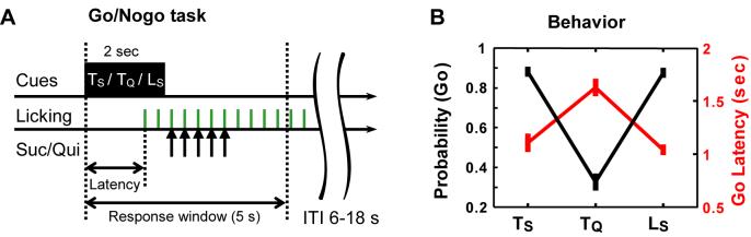
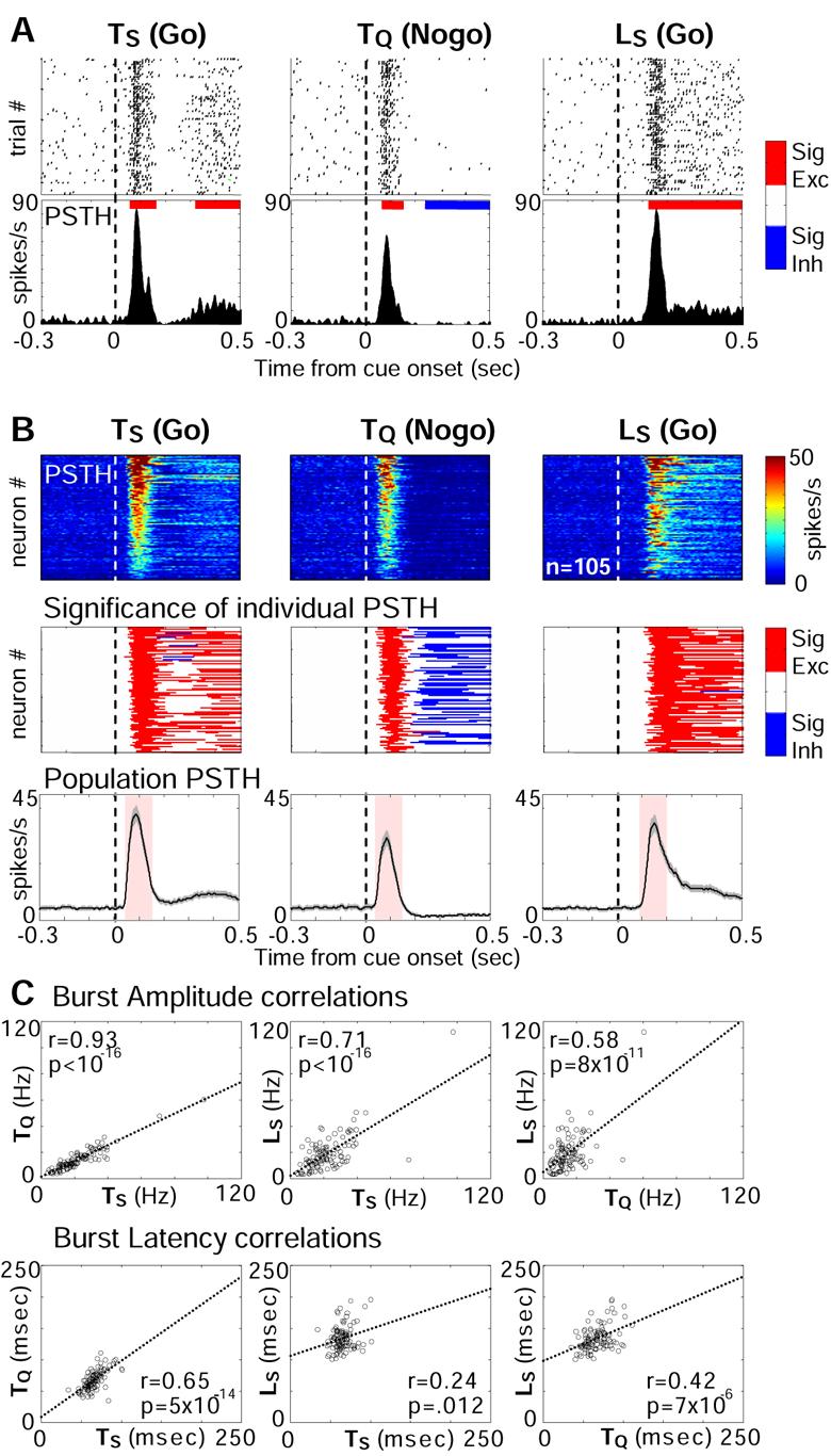
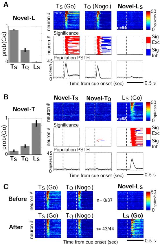
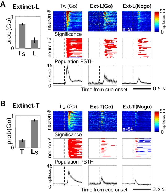
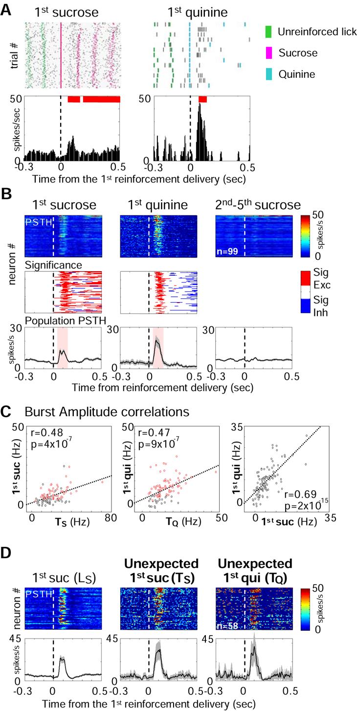
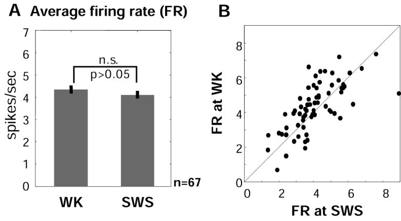
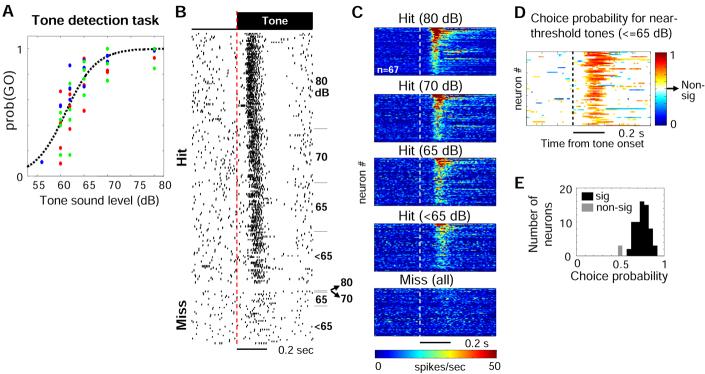
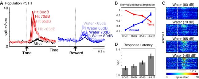
Both reward- and punishment-related stimuli are motivationally salient and attract the attention of animals. However, it remains unclear how motivational salience is processed in the brain. Here, we show that both reward- and punishment-predicting stimuli elicited robust bursting of many noncholinergic basal forebrain (BF) neurons in behaving rats. The same BF neurons also responded with similar bursting to primary reinforcement of both valences. Reinforcement responses were modulated by expectation, with surprising reinforcement eliciting stronger BF bursting. We further demonstrate that BF burst firing predicted successful detection of near-threshold stimuli. Together, our results point to the existence of a salience-encoding system independent of stimulus valence. We propose that the encoding of motivational salience by ensemble bursting of noncholinergic BF neurons may improve behavioral performance by affecting the activity of widespread cortical circuits and therefore represents a novel candidate mechanism for top-down attention.


Figures

Figure 1
Figure 1. The Go/Nogo task
(A) Schematic of the Go/Nogo task. In each trial, one of three cues, TS, TQ or LS (T, tone; L, light; subscripts S/Q indicate the associated sucrose or quinine reinforcement) was randomly chosen and presented for 2 seconds. Licking within the 5-sec response window lead to delivery of the corresponding reinforcement at the 3rd-7th licks. Licking outside the response window lead to reset of the inter-trial interval (ITI) counter. Latency, time to first lick. (B) Behavioral performance in the Go/Nogo task. The probability of Go responses (black line, left-axis) and the average latency for Go responses (red line, right-axis) for each cue (mean ± s.e.m, n=4 rats, 19 sessions). Notice that for TQ trials, the correct behavioral response is Nogo.
Figure 2
Figure 2. Motivationally salient cues elicit bursting responses of BF neurons
(A) Bursting responses of one BF neuron to cues when the rat made correct behavioral responses. Upper panels, raster plots aligned to cue onsets. The response latency in all trials exceeded 0.5 sec and thus was not shown. Lower panels, peri-stimulus time histogram (PSTH) of the same responses. The red and blue lines on top of the PSTHs indicated significant excitatory and inhibitory responses (p=0.005), respectively, calculated based on [−1,0] sec baseline PSTH before cue onsets. (B) BF population bursting responses to cues. Upper panels, each row represented the color-coded PSTH for one neuron. Only neurons with bursting responses to all three cues were plotted, and sorted by their burst amplitude toward TS for all subplots. Middle panels, each row represented the color-coded response significance of individual PSTHs. Lower panels, average population response to the cues (mean ± s.e.m). Pink shaded areas indicated the time windows used to calculate burst amplitude (see Experimental Procedures). (C) Correlations of burst amplitude and latency to each cue. Each circle represented one BF neuron from Figure 2B. Dotted lines were the best linear fit. The Pearson correlation coefficient and the p-values were labeled for each plot.
Figure 3
Figure 3. Absence of BF bursting responses to novel cues before learning cue-reinforcement associations
(A) Behavioral and neuronal responses to cues in novel-L rats, which had never experienced LS during training. BF neurons with bursting responses to both TS and TQ were plotted, and sorted by their burst amplitude to TS. Conventions as in Figure 2B. Notice the lack of behavioral and neuronal responses to the novel cue (LS), in contrast to prominent bursting responses to learned cues (TS and TQ). (B) Similar results for novel-T rats. (C) BF neurons recorded in one rat before (upper) and after (lower) learning LS-sucrose association. The fractions indicated the proportion of neurons with bursting response to both TS and TQ that also showed bursting response to LS.
Figure 4
Figure 4. Diminished BF bursting responses to cues after extinction
(A) Behavioral and neuronal responses to cues in extinct-L rats that underwent extinction training of the LS-sucrose association. Neurons with bursting responses to TS were plotted. Conventions as in Figure 2B. Responses to LS were plotted separately by rats' behavior responses (Go vs. Nogo). Notice the diminished neuronal responses to the extinguished cue (LS), in contrast to prominent bursting responses to the rewarded cue (TS). (B) Similar results for extinct-T rats.
Figure 5
Figure 5. Bursting responses of BF neurons to sucrose and quinine
(A) Responses of the BF neuron in Figure 2A to the first delivery of sucrose or quinine in each trial. Notice that rats had to lick twice (unreinforced) before sucrose or quinine was delivered. (B) BF population bursting responses to sucrose and quinine. BF neurons with bursting responses to all three cues in Figure 2B and with at least 10 quinine trials were plotted (n=99). Significant responses were calculated based on [−1,0] sec baseline PSTH prior to cue onsets. Notice that bursting response was present only to the first, but not to subsequent 2nd-5th delivery of sucrose, nor to unreinforced licks. Pink shaded areas indicated the time windows used to calculate burst amplitude. (C) Correlations of burst amplitude to sucrose, quinine and to cues, as indicated in each plot. Red circles in left and middle panels represented neurons with significant bursting responses to sucrose or quinine. (D) BF population bursting responses to sucrose and quinine from neurons in the Novel-T group (Figure 3B). Notice that bursting responses to unexpected deliveries of sucrose and quinine following novel cues, TS and TQ (middle and right), were robust at the population level. The trial numbers were low because rats had never learned to associate TS and TQ with the delivery of tastants during training,
Figure 6
Figure 6. Salience-encoding BF neurons do not change average firing rates between waking and slow-wave sleep
(A) Average firing rates of BF neurons (from Figure 2B) in the WK and SWS states (mean ± s.e.m.) were statistically not different. Only neurons with at least 10 minutes of SWS recording (n=67) were included in the calculation. (B) Scatter plot of the average firing rate at WK vs. SWS states. Each dot represented one BF neuron. The dashed line indicated equivalent firing rate between the two states.
Figure 7
Figure 7. BF bursting responses predict successful detection of near-threshold tones
(A) Probability of detecting tones at various sound pressure levels against a 61dB background noise. Colored-dots represented data from three different rats. The dashed line indicated the logistic fit. (B) Response of a BF neuron to tone onsets, sorted by hit and miss trials (left), and then by tone sound levels (right). Bursting responses were present for hit trials but not for miss trials, regardless of tone sound levels. (C) Population responses to tone onsets in hit and miss trials for neurons with bursting responses to the 80dB tone. (D) Neuronal discrimination between hit and miss trials for near-threshold tones (≦65 dB), calculated according to signal detection theory. Each row represented the color-coded choice probability of a BF neuron from Figure 7C as a function of time (see Experimental Procedures). Only bins reaching statistical significance were plotted. (E) Distribution of the maximal choice probability in the [0.05 0.25] sec interval. 64/67 neurons in Figure 7E reached significance level.
Figure 8
Figure 8. Graded BF bursting responses in the detection task
(A) Average population response to the tones and reward delivery in the detection task, sorted by tone sound levels. Notice that stronger bursting responses to target tones were associated with weaker responses to reward delivery, and vise versa. (B) Normalized burst amplitude to tones (red) and reward delivery (blue) (mean ± s.e.m, n=67). Burst amplitude of each BF neuron at each condition was normalized to its burst amplitude toward 80 dB tone (hit trials). (C) Population responses to the water reward for the same BF neurons as in Figure 7C. Notice that the bursting response is temporally broader than those in Figure 5, which likely reflects the slight temporal jitter between nose poke and licking for water reward in this experiment. (D) Mean behavioral response latency in hit trials, sorted by tone sound levels (mean ± s.e.m, n=3 rats, 11 sessions). Notice that stronger bursting responses to target tones were associated with faster response latencies.

Comment in

References

    1. Alonso A, Khateb A, Fort P, Jones BE, Muhlethaler M. Differential oscillatory properties of cholinergic and noncholinergic nucleus basalis neurons in guinea pig brain slice. Eur J Neurosci. 1996;8:169–182. - PubMed
    1. Aston-Jones G, Cohen JD. An integrative theory of locus coeruleus-norepinephrine function: adaptive gain and optimal performance. Annu Rev Neurosci. 2005;28:403–450. - PubMed
    1. Belova MA, Paton JJ, Morrison SE, Salzman CD. Expectation modulates neural responses to pleasant and aversive stimuli in primate amygdala. Neuron. 2007;55:970–984. - PMC - PubMed
    1. Burk JA, Sarter M. Dissociation between the attentional functions mediated via basal forebrain cholinergic and GABAergic neurons. Neuroscience. 2001;105:899–909. - PubMed
    1. Buzsaki G, Bickford R, Ponomareff G, Thal L, Mandel R, Gage F. Nucleus basalis and thalamic control of neocortical activity in the freely moving rat. J. Neurosci. 1988;8:4007–4026. - PMC - PubMed

Publication types