. 2008 Jul 11;31(1):124-33.
doi: 10.1016/j.molcel.2008.06.011.

Structural basis for the autoinhibition of talin in regulating integrin activation

Affiliations

Structural basis for the autoinhibition of talin in regulating integrin activation

Esen Goksoy et al. Mol Cell. .

Abstract

Activation of heterodimeric (alpha/beta) integrin transmembrane receptors by the 270 kDa cytoskeletal protein talin is essential for many important cell adhesive and physiological responses. A key step in this process involves interaction of phosphotyrosine-binding (PTB) domain in the N-terminal head of talin (talin-H) with integrin beta membrane-proximal cytoplasmic tails (beta-MP-CTs). Compared to talin-H, intact talin exhibits low potency in inducing integrin activation. Using NMR spectroscopy, we show that the large C-terminal rod domain of talin (talin-R) interacts with talin-H and allosterically restrains talin in a closed conformation. We further demonstrate that talin-R specifically masks a region in talin-PTB where integrin beta-MP-CT binds and competes with it for binding to talin-PTB. The inhibitory interaction is disrupted by a constitutively activating mutation (M319A) or by phosphatidylinositol 4,5-bisphosphate, a known talin activator. These data define a distinct autoinhibition mechanism for talin and suggest how it controls integrin activation and cell adhesion.


Figures
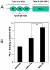
Figure 1
Figure 1
Effects of full length talin and talin-H on integrin activation. (A) Domain organization of full length talin. The three lobes of talin-H FERM domain, F1, F2, F3 or PTB and the rod domain have been labeled. (B) Comparison of the activation of integrin αIIbβ3 by full-length talin and talin-H. CHO cells stably expressing the integrin αIIbβ3 were transfected with cDNAs for full-length talin or talin-H, each as EGFP constructs. The EGFP positive cells were gated and used to monitor PAC-1 binding to determine the extent of αIIbβ3 activation as previously described (Ma et al 2008,). The data are means ± S.D. from three independent experiments. **P<0.01.
Figure 2
Figure 2
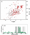
The binding of talin-RM to talin-F2F3. (A). 2D TROSY 1H-15N HSQC of talin-F2F3 in the absence (black) and presence of unlabeled talin-RM (red). Well resolved peaks, which are either significantly broadened or shifted, are labeled in free form talin-F2F3. (B) Chemical shift mapping of the binding to talin-F2F3. Only the resonances in F3 (306-405) are significantly perturbed. Residues whose signals were diminished due to severe line-broadening are indicated by grey bars.
Figure 3
Figure 3
Mapping of the talin-RM binding site on talin-PTB. (A) 1H/15N Chemical shift changes of talin-PTB upon binding to talin-RM as a function of residue number. As a comparison, the chemical shift changes of talin-PTB upon binding to β3 CT chimera is also reproduced using the chemical shift table deposited in the BioMagResBank (accession number 7150) by Wegener et al., 2007. The membrane-distal residues involved in binding to integrin β3 CT chimera are colored in red in the upper panel. Note that W359 is changed dramatically by β3 CT chimera (the change was so big due to its bulky interaction with talin-PTB (Wegener et al., 2007). that a broken line was used to conserve space). (B). Significantly perturbed residues on talin-PTB by β3-CT chimera (left) and talin-RM (right) are highlighted using the structure of talin-PTB. The order of dark blue, blue, and light blue indicate the extent of the chemical shift changes with the dark blue having the most significant changes. The changes have some similarity indicating potential overlapping binding sites for β3 CT and talin-RM but there are significant differences indicating the binding sites are not identical.
Figure 4
Figure 4
Chemical shift perturbation profiles for WT talin-PTB and its mutants Q381V and S365D by talin-RM. Note that the chemical shifts are attenuated with the Q381V mutant but are still similar to WT, whereas they are dramatically reduced with the S365D mutant.
Figure 5
Figure 5
Talin autoinhibition and its effect on integrin activation. (A) Surface representation of talin-PTB domain with the talin-RM binding site highlighted. Residues whose mutations impair talin-RM binding are colored in green whereas other potential binding residues are colored in cyan based on their significant chemical shift perturbation in Figure 3 and competition data in Figure S6-S9. (B) The same view as (A) but with the integrin β3 CT binding site highlighted. The integrin binding site is based on Garcia-Alvarez et al., 2003 and Wegener et al., 2007. The integrin membrane-proximal β3 CT binding residues are colored in green and the membrane-distal β3 CT binding residues are colored in red. Note the significant overlap of the integrin β3 membrane-proximal binding site in green as compared to that of talin-RM in (A) also in green. (C). Comparison of the activity of integrin αIIbβ3 by full-length talin, full length talin M319A, and talin-H activation. M319A is more potent than talin-H probably due to its higher affinity for integrin β3 CT than talin-H arising from its two sites of interaction, talin-H/integrin (unmasked) and talin-R/integrin. **P<0.01; *P<0.05.(D) The same view of (A) but with potential PIP2 binding site highlighted based on the chemical shift mapping of significantly perturbed residues (Green, most significantly shifted, Cyan, next significantly shifted).
Figure 6
Figure 6
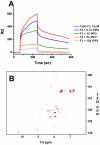
PIP2 disrupts the inhibitory talin-PTB/talin-RM interaction. (A) SPR showing that C8-PIP2 (echelon Bioscience, Inc) suppresses the talin-PTB interaction with talin-RM in a concentration dependent manner. Talin-RM was immobilized on the activated chip. Talin-PTB mixed with increasing amount of PIP2 was each injected at a flow rate of 20μl/min. (B). Overlay of HSQC spectra of 15N-labeled talin-PTB showing that C4-PIP2 (soluble at high concentration in 150mM NaCl, 50mM phosphate buffer, pH 7.0) recovers many signals of talin-PTB (red), which were otherwise broadened/disappeared in the presence of talin-RM (black) (talin-PTB:talin-RM:PIP2 = 0.2mM:0.4mM:2.0mM).
Figure 7
Figure 7
Model for the talin activation in inducing the integrin activation. In the closed state, the integrin membrane-proximal β CT binding site (blue bar) on talin-H is masked by talin-R, although talin can still weakly bind to integrin β CT via unmasked sites. Upon activation by some cellular factor such as PIP2 or RIAM, talin undergoes conformational change so that talin-PTB can access to the integrin membrane-proximal β CT and induces integrin α/β CT unclasping and integrin activation. The ternary complex involving PIP2/talin/integrin has been indicated by Martel et al., 2001 and Cluzel et al., 2005.

References

    1. Bax A, Grzesiek S. Methodological advances in protein NMR. Accounts Chem. Res. 1993;26:131–138.
    1. Bompard G, Martin M, Roy C, Vignon F, Freiss G. Membrane targeting of protein tyrosine phosphatase PTPL1 through its FERM domain via binding to phosphatidylinositol 4,5-biphosphate. J Cell Sci. 2003;116(Pt 12):2519–30. - PubMed
    1. Bonvin AM, Boelens R, Kaptein R. NMR analysis of protein interactions. Curr Opin Chem Biol. 2005;9(5):501–8. - PubMed
    1. Bouaouina M, Lad Y, Calderwood DA. The N-terminal domains of talin co-operate with the PTB-like domain to activate beta 1 and beta 3 integrins. J Biol Chem. 2007 Epub ahead of print. - PubMed
    1. Bryson K, McGuffin LJ, Marsden RL, Ward JJ, Sodhi JS, Jones DT. Protein structure prediction servers at University College London. Nucleic Acids Res. 2005;33:W36–8. Web Server issue. - PMC - PubMed

Publication types