. 2008 Sep 26;283(39):26694-704.
doi: 10.1074/jbc.M803514200. Epub 2008 Jul 8.

Structural and functional analysis of the human HDAC4 catalytic domain reveals a regulatory structural zinc-binding domain

Affiliations

Structural and functional analysis of the human HDAC4 catalytic domain reveals a regulatory structural zinc-binding domain

Matthew J Bottomley et al. J Biol Chem. .

Abstract

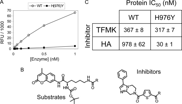
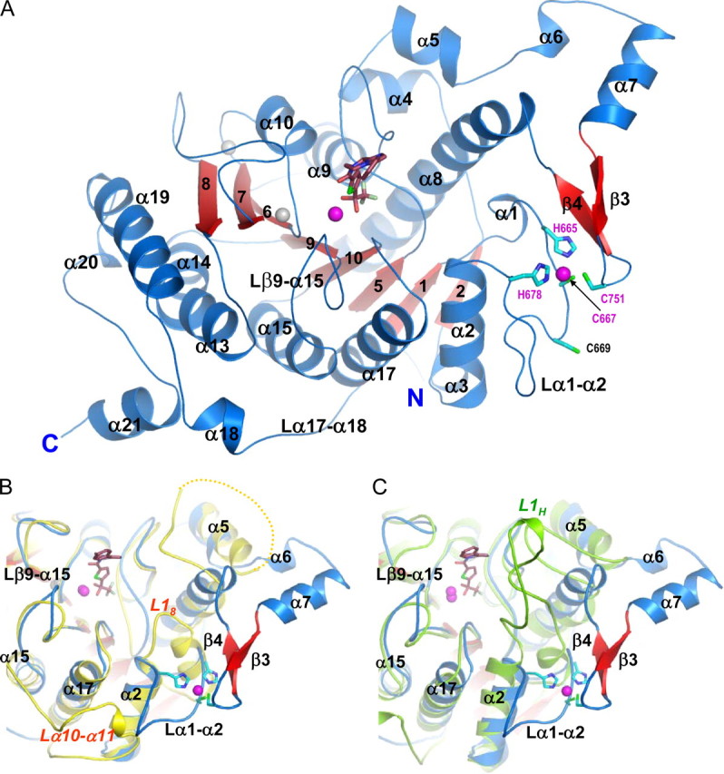
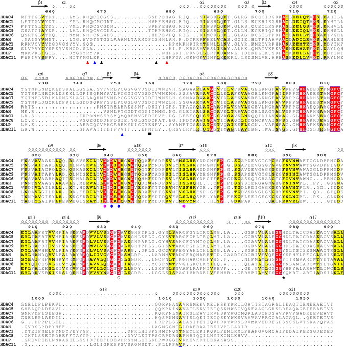
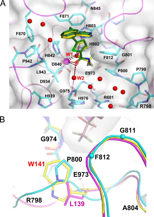
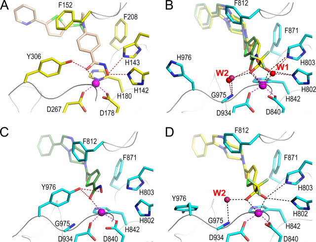
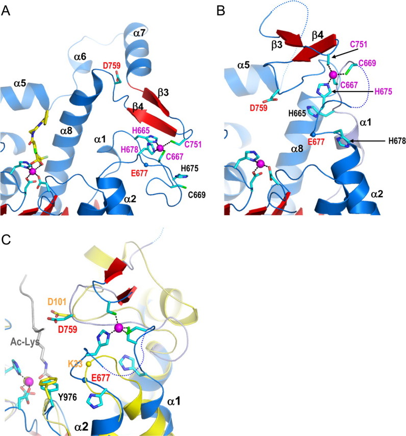
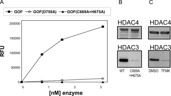
Histone deacetylases (HDACs) regulate chromatin status and gene expression, and their inhibition is of significant therapeutic interest. To date, no biological substrate for class IIa HDACs has been identified, and only low activity on acetylated lysines has been demonstrated. Here, we describe inhibitor-bound and inhibitor-free structures of the histone deacetylase-4 catalytic domain (HDAC4cd) and of an HDAC4cd active site mutant with enhanced enzymatic activity toward acetylated lysines. The structures presented, coupled with activity data, provide the molecular basis for the intrinsically low enzymatic activity of class IIa HDACs toward acetylated lysines and reveal active site features that may guide the design of class-specific inhibitors. In addition, these structures reveal a conformationally flexible structural zinc-binding domain conserved in all class IIa enzymes. Importantly, either the mutation of residues coordinating the structural zinc ion or the binding of a class IIa selective inhibitor prevented the association of HDAC4 with the N-CoR.HDAC3 repressor complex. Together, these data suggest a key role of the structural zinc-binding domain in the regulation of class IIa HDAC functions.


Figures

FIGURE 1.
FIGURE 1.
Catalytic activity and inhibition of HDAC4 wild type and mutants. A, the full-length WT HDAC4 is highly active on the trifluoroacetamide substrate, whereas the H976Y mutant is inactive. RFU, relative fluorescence units. B, top, the substrate, comprising an acetylated lysine side chain (where R represents CH3) or the trifluoroacetamide version (where R represents CF3). Bottom, the inhibitor; R represents CF3 for the trifluoromethylketone (TFMK) or NH-OH for the hydroxamic acid (HA). C, potency of TFMK and HA inhibitors for WT and GOF HDAC4cd proteins.
FIGURE 2.
FIGURE 2.
The inhibitor-bound HDAC4 catalytic domain. A, HDAC4cd bound to the TFMK inhibitor (sticks); Protein Data Bank code 2VQJ. Zinc and potassium ions are shown as magenta and gray spheres, respectively. The four residues coordinating the structural zinc ion are shown (sticks). B, a superposition of TFMK-bound HDAC4cd (blue/red) and HDAC8 (yellow). Each α-helix and β-strand of HDAC4 has a structural counterpart in HDAC8, except for α6, α7, β3, β4, and α18. The C terminus of HDAC8 coincides with the end of α19, such that α20 and α21 are also absent from HDAC8. Loop L18 of HDAC8 is much shorter than loop α1-α2 of HDAC4. The yellow dotted line indicates HDAC8 residues missing due to disorder. C, a superposition of TFMK-bound HDAC4cd (blue/red) and HDAH (green). Loop L1H of HDAH is much longer than loop α1-α2 of HDAC4. Both L1H and L18 loops turn inward toward the catalytic site. The major differences with HDAH occur in this loop and in the α6-α7-β3-β4 region and in the longer C terminus of HDAC4.
FIGURE 3.
FIGURE 3.
Multiple sequence alignment of HDAC catalytic domains. Shown is a structure-based multiple sequence alignment, made using the ESPript program, of HDACs from classes I, IIa, IIb, and IV and the bacterial HDAC-like proteins, HDLP and HDAH. Secondary structure elements are shown for the TFMK-bound HDAC4cd structure; numbering is for wild type HDAC4. Blue triangles, binds structural zinc ion in inhibited and inhibitor-free structures; red triangles, only binds structural zinc ion in the inhibited protein; black triangles, only binds structural zinc ion in the inhibitor-free protein; blue circles, binds both the catalytic zinc ion (via side chains) and potassium ion-1 (via backbone); open circle, binds the catalytic zinc ion (via side chain); pink circles, binds only potassium ion-1 via side chains (Asp838 and Ser861) or backbone (Leu862); black star, “gain-of-function” residue; black square, mutation to Ala reduces activity.
FIGURE 4.
FIGURE 4.
Interactions of HDAC4cd with inhibitors. A, interactions of the TFMK (yellow carbons) and HA (green carbons) with HDAC4; the complex structures have PDB codes 2VQJ and 2VQM, respectively. Red spheres, water molecules. The surface around the protein is shown for the TFMK-bound HDAC4cd. B, superposition of HDAC4cd (cyan) bound to TFMK (sticks and surface) with HDAC8 (yellow) and homology-modeled HDAC1 (magenta). Residues surrounding the trifluoro group are labeled. Cyan spheres, Cα atoms.
FIGURE 5.
FIGURE 5.
The active sites of HDAC4 and HDAC8. A, the active site of HDAC8 (yellow side chains) bound to a hydroxamic acid inhibitor (light brown) from Protein Data Bank entry 1W22. B, WT HDAC4cd with bound TFMK (yellow carbons) (Protein Data Bank code 2VQQ) and superposed HA (green carbons) (Protein Data Bank code 2VQM). The active site closely resembles HDAC8. C, the active site of GOF HDAC4cd with HA bound (Protein Data Bank code 2VQV); Tyr976 adopts the inward, class I-like conformation. D, the active site of GOF HDAC4cd with TFMK bound (Protein Data Bank code 2VQO); Tyr976 adopts an outward conformation.
FIGURE 6.
FIGURE 6.
Conformational flexibility of the structural zinc-binding domain and role in substrate binding. A, the active site of TFMK-bound WT HDAC4cd showing the original structural zinc-binding scheme (cyan sticks). B, the new conformation of the zinc-binding region in the inhibitor-free GOF HDAC4cd structure (Protein Data Bank code 2VQW). C, a structural comparison of the active sites of the inhibitor-free GOF HDAC4cd structure (blue/red) and HDAC8 (yellow) bound to an acetylated lysine peptidic substrate (gray). The blue and yellow spheres representing the Cα atoms of HDAC4 Glu677 and HDAC8 Lys33, respectively, illustrate how this inhibitor-free HDAC4 structure has a more closed active site due to the new conformation of the α1-α2 loop.
FIGURE 7.
FIGURE 7.
Role of the HDAC4 structural zinc-binding domain. A, activities on the acetamide substrate of the GOF HDAC4cd protein, the GOF HDAC4cd D759A mutant, and the GOF HDAC4cd C669A/H675A mutant. B, full-length HDAC4 proteins (WT and C669A/H675A mutant; top) were immunopurified and probed by Western blotting for HDAC3 (bottom). HDAC3 did not co-purify with the mutant protein. C, HDAC3 did not co-purify with full-length WT HDAC4 when expressed in the presence of a cell-permeable class IIa-specific trifluoromethylketone inhibitor, as shown by Western blotting.

References

    1. Cress, W. D., and Seto, E. (2000) J. Cell Physiol. 184 1-16 - PubMed
    1. Ito, A., Kawaguchi, Y., Lai, C. H., Kovacs, J. J., Higashimoto, Y., Appella, E., and Yao, T. P. (2002) EMBO J. 21 6236-6245 - PMC - PubMed
    1. Glozak, M. A., Sengupta, N., Zhang, X., and Seto, E. (2005) Gene (Amst.) 363 15-23 - PubMed
    1. Chen, L., Fischle, W., Verdin, E., and Greene, W. C. (2001) Science 293 1653-1657 - PubMed
    1. Yuan, Z. L., Guan, Y. J., Chatterjee, D., and Chin, Y. E. (2005) Science 307 269-273 - PubMed