. 2008 Sep 12;283(37):25428-25436.
doi: 10.1074/jbc.M804146200. Epub 2008 Jul 9.

Identification and functional characterization of adipose-specific phospholipase A2 (AdPLA)

Affiliations

Identification and functional characterization of adipose-specific phospholipase A2 (AdPLA)

Robin E Duncan et al. J Biol Chem. .

Abstract

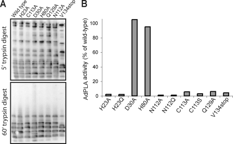
Phospholipases A(2) (PLA(2)s) catalyze hydrolysis of fatty acids from the sn-2 position of phospholipids. Here we report the identification and characterization of a membrane-associated intracellular calcium-dependent, adipose-specific PLA(2) that we named AdPLA (adipose-specific phospholipase A(2)). We found that AdPLA was highly expressed specifically in white adipose tissue and was induced during preadipocyte differentiation into adipocytes. Clearance of AdPLA by immunoprecipitation significantly decreased PLA activity in white adipose tissue lysates but had no effect on liver lysates, where expression was hardly detectable. In characterizing AdPLA, we employed radiochemical assays with TLC analysis of the enzyme activity of lysates from COS-7 cells overexpressing AdPLA. For kinetic studies, we produced purified recombinant AdPLA for use in a lipoxidase-coupled spectrophotometric assay. AdPLA generated free fatty acid and lysophospholipid from phosphatidylcholine with a preference for hydrolysis at the sn-2 position. Although we found low but detectable lysophospholipase activity, AdPLA showed no significant activity against a variety of other lipid substrates. Calcium was found to activate AdPLA but was not essential for activity. Studies with known phospholipase inhibitors, including bromoenolactone, methyl arachidonyl fluorophosphate, AACOCF(3), 7,7-dimethyl-5,8-eicosadienoic acid, and thioetheramide, supported that AdPLA is a phospholipase. Mutational studies showed that His-23 and Cys-113 are critical for activity of AdPLA and suggested that AdPLA is likely a His/Cys PLA(2). Overall, although AdPLA is similar to other histidine phospholipases in pH and calcium dependence, AdPLA showed different characteristics in many regards, including predicted catalytic mechanism. AdPLA may therefore represent the first member of a new group of PLA(2)s, group XVI.


Figures

FIGURE 1.
FIGURE 1.
AdPLA is highly expressed in adipose tissue and adipocytes. A, AdPLA mRNA expression (left panel) was 40- to over 150-fold higher in subcutaneous (subcut), renal, and gonadal WAT depots than in liver, and 18-fold higher in brown adipose tissue (BAT)(n = 3). In contrast, cPLA2α (middle panel) and iPLA2β (right panel) were found to be expressed in a wide variety of tissues at varying levels, showing no obvious tissue-specific expression pattern. sk musc, skeletal muscle. B, AdPLA mRNA is induced during late stage differentiation of 3T3-L1 and primary preadipocytes. C, representative immunoblot showing that AdPLA protein is barely detectable in 3T3-L1 preadipocytes but is highly induced upon differentiation. D, AdPLA is induced in 3T3-L1 cells by a differentiation mixture of dexamethasone (DEX) + MIX but not by either agent alone. E, subcellular fractionation of COS-7 cells overexpressing HA-tagged AdPLA indicated localization of AdPLA both to the 100,000 × g membrane fraction as well as in the cytosolic fraction. F, confocal microscopy images showing a punctate localization of GFP-tagged full-length AdPLA predominantly in the perinuclear region of COS-7 cells. Truncated AdPLA lacking the membrane-spanning C terminus domain assumed a more diffuse localization. G, full-length AdPLA-GFP co-localized partially with COX-1 in COS-7 cells. H, full-length AdPLA-GFP showed a perinuclear localization in differentiated 3T3-L1 cells and partially co-localized with the endoplasmic reticulum (ER).
FIGURE 2.
FIGURE 2.
AdPLA has phospholipase activity but lacks acyltransferase activity. A, representative immunoblot of COS-7 cell lysates transiently transfected with HA-tagged AdPLA. B, NUS-AdPLA tagged with HA and His was affinity-purified on Co3+-agarose beads. Following SDS-PAGE, NUS-AdPLA was detectable as a single band of ∼81 kDa by Coomassie staining (lane 1, molecular weight standards; lane 2, NUS-AdPLA) and by immunoblotting on polyvinylidene difluoride with anti-HA (lane 3). C, COS-7 cell lysates overexpressing AdPLA or control vector were incubated with 1,2-di[1-14C]palmitoyl-sn-glycerol-3-phosphocholine (Sn1*2*PC) and DAG. AdPLA overexpression increased the appearance of [14C]palmitate, indicating phospholipase activity, and this effect was enhanced by increasing the incubation time or amount of protein. There was no evidence of acyltransferase activity (i.e. no 14C-TAG formed). MAG, monoacylglycerol; PL, phospholipid. D, COS-7 cell lysates overexpressing AdPLA caused a greater release of 14C-FFA from a complex mixture of radiolabeled cellular lipids, but otherwise showed no changes in any other lipid groups. E, cytosolic and membrane fractions derived from COS-7 cells transfected with AdPLA or control vector incorporated [14C]palmitoyl-CoA in a similar manner, indicating no apparent acyl-CoA-dependent acyltransferase activity. F, incubation of lysates overexpressing AdPLA with 1,2-di[1-14C]palmitoyl-sn-glycerol-3-phosphocholine (Sn1*2*PC) yielded an increase in the production of radiolabeled FFA and lysophosphatidylcholine (LPC), indicating that AdPLA is a PLA (n = 4). G, COS-7 cells overexpressing AdPLA or control vector incorporated similar amounts of [U-14C]palmitate during an initial 4-h pulse-labeling period. H, overexpression of AdPLA, however, resulted in a significantly greater release of FFA to the medium during the ensuing cold chase period, as determined by liquid scintillation counting of medium, and by extraction of lipids from medium and resolution by TLC (inset)(n = 12). I, clearance by immunoprecipitation (IP) of lysates from WAT with anti-AdPLA antiserum (1:100) decreased levels of AdPLA relative to lysates cleared with preimmune serum (1:100), as shown in a representative immunoblot. AdPLA was essentially undetectable in lysates from liver, and glyceraldehyde-3-phosphate dehydrogenase (GAPDH) was unchanged by immunoprecipitation. This decrease in immunoreactive AdPLA level in WAT lysates was associated with a significant decrease in PLA activity. No change in PLA activity was evident in lysates from liver (n = 7).
FIGURE 3.
FIGURE 3.
AdPLA is primarily a phospholipase. A, recombinant AdPLA and sPLA2 (positive control) efficiently hydrolyzed 1,2-dilinoleoyl-PC (PC). Activity of AdPLA and sPLA2 toward cholesteryl-linoleate (CE) or 1,3-dilinoleoyl-sn-2-glycerol (DAG) was not different from control levels (NUS alone) (***, p < 0.001 versus NUS). B, lysates from COS-7 cells transiently transfected with AdPLA or desnutrin were assayed for activity against radiolabeled triolein. **, p < 0.01, ***, p < 0.001, versus NUS. C, similar to other phospholipases, recombinant AdPLA displayed minor hydrolase activity toward 1-[1-14C]palmitoyl-2-hydroxy-sn-glycerol-3-lysophosphocholine (Lyso-PC), but significantly greater activity toward 1-palmitoyl-2-[1-14C]palmitoyl-sn-glycerol-3-phosphocholine (PC) under the same conditions (***, p < 0.001). All data shown are means ± S.E. (n = 3–4).
FIGURE 4.
FIGURE 4.
Characteristics of AdPLA phospholipase activity. Recombinant AdPLA was assayed in 50 mm Tris-HCl with 2 mm deoxycholate (n = 3–4). A, recombinant partially purified AdPLA was found to have a narrow pH range for activity with an optimum pH of 8.0. B, AdPLA activity against 20 μm 1-palmitoyl-2-linoleoyl-PC increased in the presence of increasing concentrations of CaCl2, and this effect was reversed by addition of a calcium chelator (3 mm EGTA). C, concentration-dependent activity of AdPLA against 1-palmitoyl-2-linoleoyl-PC at pH 8.0 with 2 mm CaCl2. Inset, double-reciprocal plot. D, regioselectivity of AdPLA. Concentration-dependent activity of NUS-AdPLA was compared with 1-palmitoyl-2-linoleoyl-PC (1-palm-2-lin-PC) versus 1,2-dilinoleoyl-PC (1,2-dilin-PC), where only hydrolyzed unsaturated fatty acids are quantitated in spectrophotometric readings. A preference for hydrolysis of fatty acids at the sn-2 position was evident, indicating that AdPLA is a PLA2.
FIGURE 5.
FIGURE 5.
Treatment of AdPLA with known phospholipase inhibitors. A and B, AdPLA and sPLA2 were inhibited in a similar manner by treatment with 7,7-dimethyl-5,8-eicosadienoic acid or AACOCF3, respectively. C, neither AdPLA nor sPLA2 was inhibited by BEL, a suicide-substrate inhibitor of GXSXG-containing lipases. D, MAFP, an inhibitor of serine and cysteine phospholipases, inhibited AdPLA but not sPLA2. E, thioetheramide inhibited sPLA2 but not AdPLA. Results are means ± S.E. from duplicate or triplicate measurements.
FIGURE 6.
FIGURE 6.
Mutational and deletional analysis of AdPLA activity. Sequence alignment indicated that the His-23/Cys-113 motif required by LRAT for catalysis is conserved in AdPLA. A, trypsin digest of recombinant mutants indicates a similar pattern of peptide generation. B, mutation of His-23 or Cys-113 caused a complete loss of activity in vitro, as did truncation of the C-terminal membrane localization domain. Mutation of other highly conserved residues, including Asn-112 and Glu-129, also abolished activity. Mutation of other conserved residues, including Asp-30 and His-80, had no effect, indicating a specific role for residues at positions 23, 112, 113, and 129 in catalytic activity.

References

    1. Schaloske, R. H., and Dennis, E. A. (2006) Biochim. Biophys. Acta 1761 1246-1259 - PubMed
    1. Yuan, C., Rieke, C. J., Rimon, G., Wingerd, B. A., and Smith, W. L. (2006) Proc. Natl. Acad. Sci. U. S. A. 103 6142-6147 - PMC - PubMed
    1. Aubert, J., Saint-Marc, P., Belmonte, N., Dani, C., Negrel, R., and Ailhaud, G. (2000) Mol. Cell. Endocrinol. 160 149-156 - PubMed
    1. Fajas, L., Miard, S., Briggs, M. R., and Auwerx, J. (2003) J. Lipid Res. 44 1652-1659 - PubMed
    1. Forman, B. M., Tontonoz, P., Chen, J., Brun, R. P., Spiegelman, B. M., and Evans, R. M. (1995) Cell 83 803-812 - PubMed

Publication types