. 2008 Sep 12;283(37):25150-25163.
doi: 10.1074/jbc.M800882200. Epub 2008 Jul 9.

Rho Family GTPase modification and dependence on CAAX motif-signaled posttranslational modification

Affiliations

Rho Family GTPase modification and dependence on CAAX motif-signaled posttranslational modification

Patrick J Roberts et al. J Biol Chem. .

Abstract

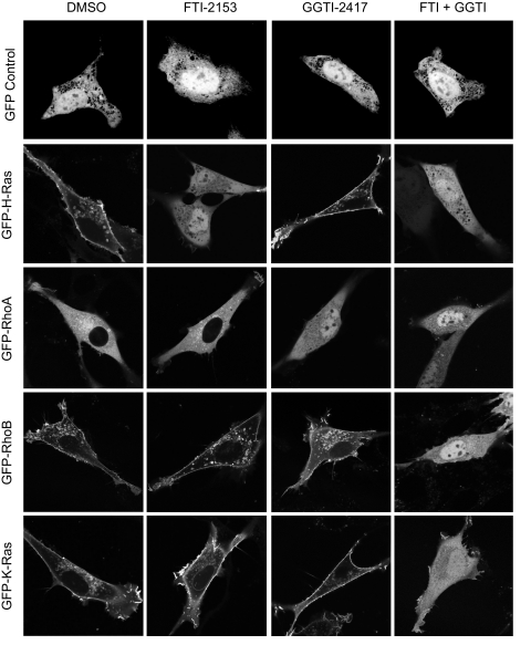
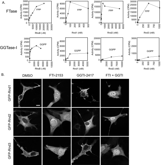
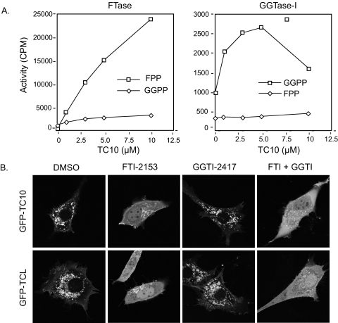
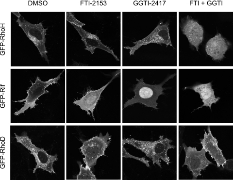
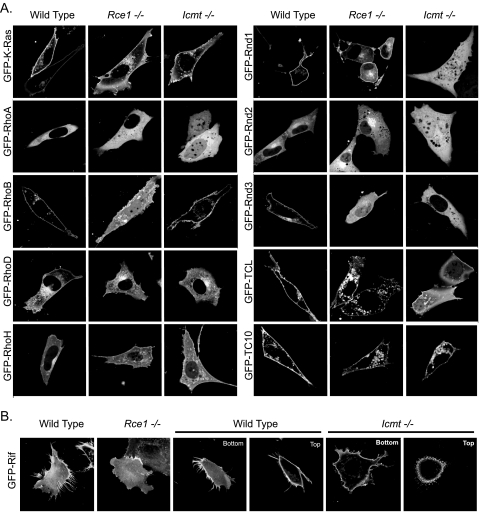
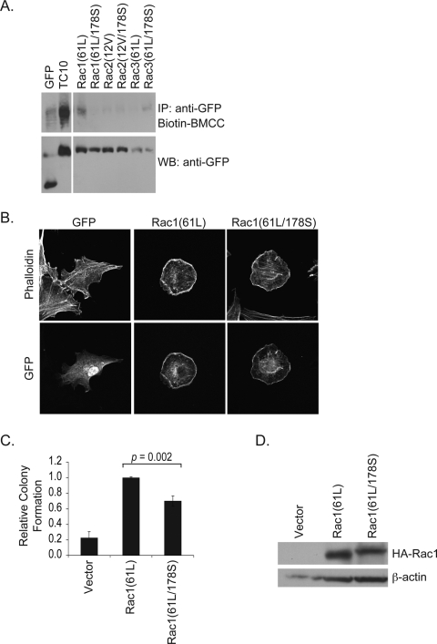
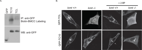
Rho GTPases (20 human members) comprise a major branch of the Ras superfamily of small GTPases, and aberrant Rho GTPase function has been implicated in oncogenesis and other human diseases. Although many of our current concepts of Rho GTPases are based on the three classical members (RhoA, Rac1, and Cdc42), recent studies have revealed the diversity of biological functions mediated by other family members. A key basis for the functional diversity of Rho GTPases is their association with distinct subcellular compartments, which is dictated in part by three posttranslational modifications signaled by their carboxyl-terminal CAAX (where C represents cysteine, A is an aliphatic amino acid, and X is a terminal amino acid) tetrapeptide motifs. CAAX motifs are substrates for the prenyltransferase-catalyzed addition of either farnesyl or geranylgeranyl isoprenoid lipids, Rce1-catalyzed endoproteolytic cleavage of the AAX amino acids, and Icmt-catalyzed carboxyl methylation of the isoprenylcysteine. We utilized pharmacologic, biochemical, and genetic approaches to determine the sequence requirements and roles of CAAX signal modifications in dictating the subcellular locations and functions of the Rho GTPase family. Although the classical Rho GTPases are modified by geranylgeranylation, we found that a majority of the other Rho GTPases are substrates for farnesyltransferase. We found that the membrane association and/or function of Rho GTPases are differentially dependent on Rce1- and Icmt-mediated modifications. Our results further delineate the sequence requirements for prenyltransferase specificity and functional roles for protein prenylation in Rho GTPase function. We conclude that a majority of Rho GTPases are targets for pharmacologic inhibitors of farnesyltransferase, Rce1, and Icmt.


Figures

FIGURE 1.
FIGURE 1.
The specific isoprenoid modifications of Ras and Rho GTPases in vivo are accurately defined using specific pharmacologic inhibitors of FTase and GGTase-I. NIH 3T3 cells were transiently transfected with expression constructs for GFP alone or GFP-tagged fusion proteins of the indicated Ras or Rho GTPases and treated with FTI-2153, GGTI-2417, or both (10 μm each) or DMSO. Live cells were visualized using confocal microscopy. Images shown are representative of three independent experiments with >80 cells examined per assay. Scale bar, 10 μm.
FIGURE 2.
FIGURE 2.
Rnd proteins are farnesylated in vitro and in vivo. A, in vitro RhoB is a strong substrate for both FTase and GGTase-I, whereas Rnd proteins are strong substrates for FTase and weak substrates for GGTase-I. Purified recombinant RhoB and Rnd proteins were added to our standard FTase or GGTase-I reaction mixture containing radiolabeled FPP or GGPP, respectively, and processed as described under “Experimental Procedures.” B, both membrane association and function of Rnd proteins are inhibited by FTI treatment. NIH 3T3 cells were transiently transfected with expression constructs for GFP-tagged fusion proteins with the indicated Rho GTPases and treated with FTI-2153, GGTI-2417, both (10 μm each), or DMSO. Live cells were visualized using confocal microscopy. Images shown are representative of three independent experiments with >80 cells examined per assay. Scale bar, 10 μm.
FIGURE 3.
FIGURE 3.
TC10 and TCL are farnesylated in vitro and in vivo. A, TC10 is a substrate for FTase and GGTase-I in vitro. Assays were done as described in the legend to Fig. 2A and under “Experimental Procedures.” B, TC10 subcellular localization is partially dependent on farnesylation. NIH 3T3 cells transfected with expression constructs for GFP-tagged fusion proteins of human TC10 or TCL were treated with FTI-2153, GGTI-2417, both (10 μm each), or DMSO. Live cells were visualized using confocal microscopy. Images shown are representative of three independent experiments with >80 cells examined per assay. Scale bar, 10 μm.
FIGURE 4.
FIGURE 4.
Atypical Rho GTPases are not simple substrates for FTase or GGTase-I. RhoH and Rif are farnesylated and/or geranylgeranylated in vivo. NIH 3T3 cells were transfected with expression constructs for the indicated GFP-tagged Rho GTPase and treated with FTI-2153, GGTI-2417, or both (10 μm each) or DMSO. Live cells were visualized using confocal microscopy. Images shown are representative of three independent experiments with >80 cells examined per assay. Scale bar, 10 μm.
FIGURE 5.
FIGURE 5.
Differential requirements for Rce1- and Icmt-mediated postprenyl processing in the subcellular localization and function of Rho family proteins. Wild-type, Rce1–/–, and Icmt–/– MEFs were transiently transfected with expression constructs for GFP-tagged fusion proteins of the indicated Rho GTPases. A, live cells were visualized using confocal microscopy. Images shown are representative of three independent experiments with >80 cells examined per assay. B, Icmt-mediated processing of Rif is not required for membrane association but contributes to subcellular membrane distribution. GFP-Rif localization was examined by Z-sectioning. The bottom and top stacks of each cell were examined for the presence of filopodia, and images shown are representative of three independent experiments with >50 cells examined per assay. Scale bar, 10 μm.
FIGURE 6.
FIGURE 6.
Distinct roles of conserved cysteines in palmitoylation and protein function of Rac. A, 293T cells were transiently transfected with expression constructs for GFP-tagged activated Rac1(61L), Rac2(12V), and Rac3(61L) with or without an additional C178S mutation; TC10; or empty vector and subjected to the biotin-BMCC labeling assay. Levels of biotin-labeled GTPases were measured by Western blotting with streptavidin-horseradish peroxidase (top). Expression of GFP-tagged constructs was confirmed by immunoblotting with anti-GFP antibodies (bottom). B, NIH 3T3 cells were transiently transfected with expression vectors encoding GFP-tagged activated Rac1(61L) or mutant Rac1(61L/178S) proteins and stained with Alexa 594-conjugated phalloidin to visualize the actin cytoskeleton. Images shown are representative of three independent experiments with >80 cells examined per assay. C, mutation of C178 causes a limited reduction in Rac transforming activity. Single cell suspensions of NIH 3T3 cells stably expressing activated Rac1 proteins were suspended in soft agar, and colony formation was monitored after 2 weeks. The number of colonies was quantified as described under “Experimental Procedures.” Colony numbers were normalized to those seen with Rac1(61L). D, expression of hemagglutinin (HA)-tagged Rac constructs was confirmed by immunoblotting with anti-hemagglutinin antibodies. IP, immunoprecipitation; WB, Western blot.
FIGURE 7.
FIGURE 7.
Methylation and palmitoylation act as redundant membrane targeting signals for TC10 localization. A, 293T cells were transiently transfected with expression constructs for GFP-tagged TC10, TCL, or empty vector and subjected to the biotin-BMCC labeling assay. Levels of biotin-labeled GTPases were measured by Western blotting with streptavidin-horseradish peroxidase (top). Expression of GFP-tagged constructs was confirmed by immunoblotting with anti-GFP antibodies (bottom). B, wild-type or Icmt–/– MEFs were transiently transfected with expression constructs for the indicated GFP-tagged Rho GTPase. Cells expressing GFP-TCL and GFP-TC10 were treated with 100 μm 2-BP and imaged alive. Images shown are representative of three independent experiments with >80 cells examined per assay. Scale bar, 10 μm. IP, immunoprecipitation; WB, Western blot.

References

    1. Colicelli, J. (2004) Science's STKE 2004, RE13 - PMC - PubMed
    1. Wennerberg, K., Rossman, K. L., and Der, C. J. (2005) J. Cell Sci. 118 843–846 - PubMed
    1. Wennerberg, K., and Der, C. J. (2004) J. Cell Sci. 117 1301–1312 - PubMed
    1. Schmidt, A., and Hall, A. (2002) Genes Dev. 16 1587–1609 - PubMed
    1. Bernards, A., and Settleman, J. (2004) Trends Cell Biol. 14 377–385 - PubMed

Publication types