. 2008 Sep;82(18):9035-42.
doi: 10.1128/JVI.00674-08. Epub 2008 Jul 9.

Common marmosets (Callithrix jacchus) as a nonhuman primate model to assess the virulence of eastern equine encephalitis virus strains

Affiliations

Common marmosets (Callithrix jacchus) as a nonhuman primate model to assess the virulence of eastern equine encephalitis virus strains

A Paige Adams et al. J Virol. 2008 Sep.

Abstract

Eastern equine encephalitis virus (EEEV) produces the most severe human arboviral disease in North America (NA) and is a potential biological weapon. However, genetically and antigenically distinct strains from South America (SA) have seldom been associated with human disease or mortality despite serological evidence of infection. Because mice and other small rodents do not respond differently to the NA versus SA viruses like humans, we tested common marmosets (Callithrix jacchus) by using intranasal infection and monitoring for weight loss, fever, anorexia, depression, and neurologic signs. The NA EEEV-infected animals either died or were euthanized on day 4 or 5 after infection due to anorexia and neurologic signs, but the SA EEEV-infected animals remained healthy and survived. The SA EEEV-infected animals developed peak viremia titers of 2.8 to 3.1 log(10) PFU/ml on day 2 or 4 after infection, but there was no detectable viremia in the NA EEEV-infected animals. In contrast, virus was detected in the brain, liver, and muscle of the NA EEEV-infected animals at the time of euthanasia or death. Similar to the brain lesions described for human EEE, the NA EEEV-infected animals developed meningoencephalitis in the cerebral cortex with some perivascular hemorrhages. The findings of this study identify the common marmoset as a useful model of human EEE for testing antiviral drugs and vaccine candidates and highlight their potential for corroborating epidemiological evidence that some, if not all, SA EEEV strains are attenuated for humans.


Figures
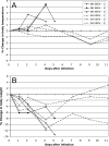
FIG. 1.
FIG. 1.
Change in body temperatures and weights of marmosets after i.n. infection with EEEV. Percent changes from the starting (day 0) body temperatures (A; in degrees Celsius) and weights (B; in grams) of marmosets were determined on days 1, 2, 4, 7, 9, and 11 after i.n. infection with either NA EEEV strain FL93-939 (solid lines) or SA EEEV strain 436087 (dashed lines). NA EEEV-1, -2, and -3 and SA EEEV-1, -2, and -3 are individual marmosets within each cohort.
FIG. 2.
FIG. 2.
Peripheral blood leukocyte subpopulations after exposure to EEEV. Marmosets were bled on days 0, 1, 2, 4, 7, 9, and 11 after i.n. infection with either NA EEEV strain FL93-939 (solid lines) or SA EEEV strain 436087 (dashed lines) to assess changes in peripheral blood leukocytes. Graphs show cell counts for total white blood cells (A), lymphocytes (B), monocytes (C), and granulocytes (D) for each marmoset. As indicated by the boxes on each graph, normal cell counts of white blood cells, lymphocytes, monocytes, and granulocytes in the peripheral blood of marmosets are 1.8 to 8.1, 0.7 to 5.0, 0.0 to 0.6, and 0.0 to 4.0 m/mm3, respectively. NA EEEV-1, -2, and -3 and SA EEEV-1, -2, and -3 are individual marmosets within each cohort.
FIG. 3.
FIG. 3.
Viremia in marmosets after i.n. infection (106 PFU) with either NA EEEV strain FL93-939 (closed circles; below limit of detection) or SA EEEV strain 436087 (open circles) (three marmosets per cohort). On day 7 after infection, one of two marmosets tested had detectable viremia (a). Error bars indicate the standard deviations, and the limit of detection by the assay was 1.7 log10 PFU/ml.
FIG. 4.
FIG. 4.
Virus titers in tissues of marmosets on days 4 and 5 after i.n. infection with NA EEEV. Marmosets were infected i.n. (106 PFU) with NA strain FL93-939. At the time of euthanasia or death (day 4 or 5 after infection), brain, lung, heart, liver, adrenal gland, lymph node, and skeletal muscle samples were collected and virus titers were determined by plaque assay. Error bars indicate the standard deviations, and the limit of detection by the assay was 1.7 log10 PFU/ml.
FIG. 5.
FIG. 5.
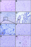
Histopathology and immunohistochemistry of marmoset brains after i.n. infection with EEEV. Similar to uninfected brains (A; magnification, ×20), there were no apparent lesions associated with SA EEEV (B; magnification, ×20); however, NA EEEV-infected marmosets showed multifocal perivascular cuffing and encephalitis in the cerebral cortex (C; magnification, ×10 [inset enlarged in panel E; magnification, ×40]) and a moderate degree of mononuclear cell leptomeningitis (D; magnification, ×40) with some perivascular hemorrhages (not shown). Similar to uninfected brains (F; magnification, ×20), SA EEEV-infected brains (G; magnification, ×20) were negative for viral antigen while NA EEEV-infected brains showed strong cytoplasmic staining (dark red) of neurons in the cerebral cortex (H; magnification, ×20). BV, blood vessel.

References

    1. Aguilar, P. V., S. Paessler, A. S. Carrara, S. Baron, J. Poast, E. Wang, A. C. Moncayo, M. Anishchenko, D. Watts, R. B. Tesh, and S. C. Weaver. 2005. Variation in interferon sensitivity and induction among strains of eastern equine encephalitis virus. J. Virol. 7911300-11310. - PMC - PubMed
    1. Aguilar, P. V., R. M. Robich, M. J. Turell, M. L. O'Guinn, T. A. Klein, A. Huaman, C. Guevara, Z. Rios, R. B. Tesh, D. M. Watts, J. Olson, and S. C. Weaver. 2007. Endemic eastern equine encephalitis in the Amazon region of Peru. Am. J. Trop. Med. Hyg. 76293-298. - PubMed
    1. Aguilar, P. V., A. P. Adams, E. Wang, A. Carrara, M. Anishchenko, I. Frolov, and S. C. Weaver. 2008. Structural and nonstructural protein genome regions of eastern equine encephalitis virus are determinants of interferon sensitivity and murine virulence. J. Virol. 824920-4930. - PMC - PubMed
    1. Alice, F. J. 1956. Infecção humana pelo virus “leste” da encefalite equina. Bol. Inst. Biol. Bahia (Brazil) 33-9.
    1. Bastian, F. O., R. D. Wende, D. B. Singer, and R. S. Zeller. 1975. Eastern equine encephalomyelitis: histopathologic and ultrastructural changes with isolation of the virus in a human case. Am. J. Clin. Pathol. 6410-13. - PubMed

Publication types

LinkOut - more resources