. 2009 Jan;129(1):70-8.
doi: 10.1038/jid.2008.191. Epub 2008 Jul 10.

Overexpression of IL-1alpha in skin differentially modulates the immune response to scarification with vaccinia virus

Affiliations

Overexpression of IL-1alpha in skin differentially modulates the immune response to scarification with vaccinia virus

Tian Tian et al. J Invest Dermatol. 2009 Jan.

Abstract

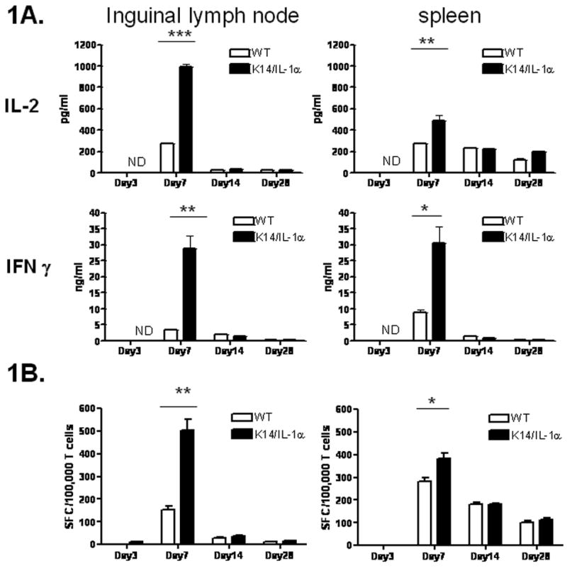
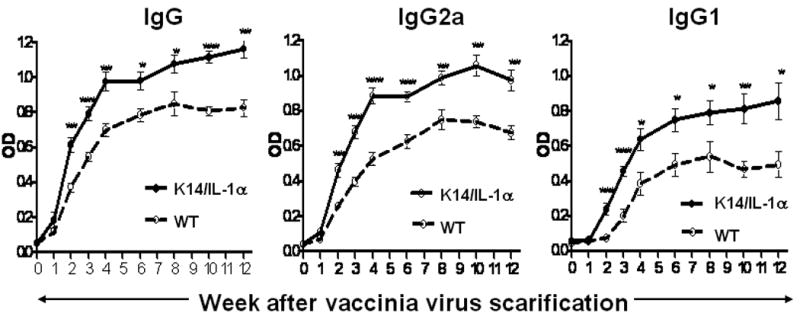
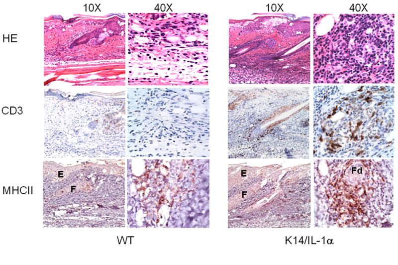
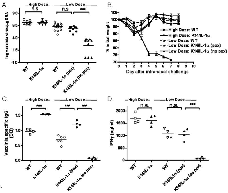
Transepidermal inoculation of vaccinia virus (VV), or scarification, has been used effectively for the induction of specific and long-lasting immunity to smallpox and is superior to other routes of immunization. Scarification of individuals with atopic skin disease or immune deficiency, however, can lead to persistent viral replication and result in significant morbidity and mortality. These effects of scarification presumably reflect the unique immunological properties of skin and the immune cells resident in, or recruited to, the site of inoculation. To explore these phenomena, we utilized transgenic mice engineered to overexpress IL-1alpha, a critical mediator of cutaneous inflammation, in the epidermis. Following scarification with VV, both transgenic and wild-type mice develop local pox. At high doses of VV, IL-1alpha transgenic mice recruited immune cells to the inoculation site more rapidly and demonstrated enhanced T-cell and humoral immune responses. At limiting doses, however, IL-1alpha transgenic mice could effectively control virus replication without formation of pox lesions or activation of a memory response. This study suggests that IL-1 might be useful as an adjuvant to enhance antiviral immunity and promote safer vaccination strategies; however, understanding the balance of IL-1 effects on innate and adaptive immune functions will be critical to achieve optimal results.


Conflict of interest statement

Conflict of Interest: The authors state no conflicts of interest.

Figures

Figure 1
Figure 1. K14/IL-1α mice develop stronger Th1 immune response following vaccinia scarification compared with WT mice
K14/IL-1α and WT mice were skin scarified at the base of their tails with 2 × 10⁶ PFU of WR-GFP vaccinia virus. At indicated time points after immunization, purified inguinal LN or splenic T cells were cultured in vitro with infected syngeneic (APC). A. IL-2 and IFN-γ in the culture supernatant measured after 40 h. Bars represent means ± SE and are representative of four independent experiments. B. Frequency of vaccinia-specific T cells measured by IFN-γ ELISPOT assay. Data are expressed as spot-forming cells (SFC) per 106 T cells. Bars represent means ± SE and are representative of two independent experiments. * = p < 0.05, ** = p < 0.01, *** = p < 0.001, ND = not detected.
Figure 2
Figure 2. Kinetics of vaccinia-specific antibody response following scarification with vaccinia virus
K14/IL-1α and WT mice were skin scarified at the base of their tails with 2 × 10⁶ PFU of WR-GFP vaccinia virus. At various time points after immunization (weekly for weeks 1 to 4 and biweekly for weeks 6 to 12), mice from each group were bled and their sera were collected. Sera were also collected from control, uninfected (week 0) mice. The kinetics of the vaccinia-specific antibody response in the sera of individual mice at each time point was measured by ELISA. Data represent means ± SE using 6 mice per group and are representative of four independent experiments. * = p < 0.05, ** = p < 0.01, *** = p < 0.001.
Figure 3
Figure 3. Histology of skin lesions from vaccinia-scarified WT and K14/IL-1α mice
Skin samples from the site of inoculation were obtained from WT and K14/IL-1α mice 3 days after scarification with 2 × 10⁶ PFU of WR-GFP vaccinia virus. Formalin-fixed paraffin-embedded tail-skin samples were stained with H&E as well as anti-CD3 and anti-Ia (MHC Class II) mAb. Labels indicate epidermis (E), proximal follicle (F), and distal follicle (Fd) regions. The data shown are representative of three to four individual mice per experimental group.
Figure 4
Figure 4. Phenotypic maturation of inguinal LN DC after vaccinia scarification
Cells recovered from inguinal lymph nodes harvested at the time points indicated were stained for CD11c and MHC Class II, CD80 or CD86. Bars represent mean fluorescence intensity of MHC Class II, CD80 and CD86 on CD11c+ gated cells. Data represent the means ± SE of four to five individual mice in one experiment. * = p < 0.05, ** = p < 0.01, *** = p < 0.001, n.s. = not significant.
Figure 5
Figure 5. Influence of low-dose vaccinia virus on protection in mouse challenge models
K14/IL-1α and WT mice were skin scarified at the base of their tails with 2 × 10⁶ PFU (high dose) or 5 × 10³ PFU (low dose) of WR-GFP vaccinia virus. All of the WT mice, immunized with either high dose (n= 10) or the low dose (n= 20) vaccinia virus, developed pox lesions. Of K14/IL-1α mice immunized with low-dose virus (n=20), half did not form pox lesions (n=10). A. Inoculated skin was removed at 7 days post-immunization and vaccinia viral DNA content determined by quantitative PCR. This method can detect levels as low as 1 virus copy/μg DNA. Data shown represent the individual values (symbols), and means (bars) of 9 to 20 mice per group in two experiments. *** = p < 0.001, n.s. = not significant. B. Five weeks after vaccination, mice received an intranasal challenge with 1 × 10⁷ PFU of vaccinia virus WR and were weighed daily. The data plotted represent the means ± SE for each group. C. Vaccinia-specific IgG in the sera of individual mice measured by ELISA one month after immunization. Data represent the means ± SE of 4 to 6 mice per group. *** = p < 0.001. D. Thirty-five days post-infection, whole splenocytes were prepared from each mouse and stimulated with in vitro–infected syngeneic APC. IFN-γ in the culture supernatant was measured after 40 h. Bars represent means ± SE for mice in each group. *** = p < 0.001, n.s. = not significant.

References

    1. Alcami A. Viral mimicry of cytokines, chemokines and their receptors. Nat Rev Immunol. 2003;3:36–50. - PubMed
    1. Alcami A, Smith GL. A soluble receptor for interleukin-1 beta encoded by vaccinia virus: a novel mechanism of virus modulation of the host response to infection. Cell. 1992;71:153–167. - PubMed
    1. Alcami A, Symons JA, Collins PD, Williams TJ, Smith GL. Blockade of chemokine activity by a soluble chemokine binding protein from vaccinia virus. J Immunol. 1998;160:624–633. - PubMed
    1. Bray M. Pathogenesis and potential antiviral therapy of complications of smallpox vaccination. Antiviral research. 2003;58:101–114. - PubMed
    1. Dinarello CA. Biologic basis for interleukin-1 in disease. Blood. 1996;87:2095–2147. - PubMed

Publication types

LinkOut - more resources