. 2008 Aug 5;47(31):8195-202.
doi: 10.1021/bi800551y. Epub 2008 Jul 11.

Bacterial ApbC can bind and effectively transfer iron-sulfur clusters

Affiliations

Bacterial ApbC can bind and effectively transfer iron-sulfur clusters

Jeffrey M Boyd et al. Biochemistry. .

Abstract

The metabolism of iron-sulfur ([Fe-S]) clusters requires a complex set of machinery that is still being defined. Mutants of Salmonella enterica lacking apbC have nutritional and biochemical properties indicative of defects in [Fe-S] cluster metabolism. ApbC is a 40.8 kDa homodimeric ATPase and as purified contains little iron and no acid-labile sulfide. An [Fe-S] cluster was reconstituted on ApbC, generating a protein that bound 2 mol of Fe and 2 mol of S (2-) per ApbC monomer and had a UV-visible absorption spectrum similar to known [4Fe-4S] cluster proteins. Holo-ApbC could rapidly and effectively activate Saccharomyces cerevisiae apo-isopropylmalate isolomerase (Leu1) in vitro, a process known to require the transfer of a [4Fe-4S] cluster. Maximum activation was achieved with 2 mol of ApbC per 1 mol of apo-Leu1. This article describes the first biochemical activity of ApbC in the context of [Fe-S] cluster metabolism. The data herein support a model in which ApbC coordinates an [4Fe-4S] cluster across its dimer interface and can transfer this cluster to an apoprotein acting as an [Fe-S] cluster scaffold protein, a function recently deduced for its eukaryotic homologues.


Figures

Figure 1
Figure 1
Protein sequence alignment of Nbp35, Cfd1, and ApbC. Protein Alignments were assembled using the Clustal_W method in the Lasergene® software. Sequence of interest is boxed: the Walker A box is dashed and Cys-X-X-Cys motif solid. Cysteine residues are highlighted above the consensus sequence. Proteins listed as follows: ApbC (Salmonella enterica Serovar Typhimurium LT2), Cfd1 (Saccharomyces cerevisiae), and Nbp35 (Saccharomyces cerevisiae).
Figure 2
Figure 2
UV-visible absorption spectra of holo-ApbC shows spectral features similar to known [Fe-S] cluster proteins. of ApbC after chemical reconstitution without (solid), and with 2 mM sodium dithionite (long dash) in 50 mM Tris, pH 8.0, 150 mM NaCl. ApbC was reconstituted as outlined in Materials and Methods. The concentration of ApbC is 1.1 mg/mL for the reconstituted, and reconstituted-reduced protein samples. The Figure includes a blow-up of the spectra from 300–800 nm (inset) to view spectroscopic details.
Figure 3
Figure 3
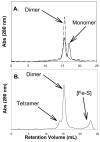
The quaternary structure of ApbC protein is in a dynamic equilibrium. Panel A: As isolated, ApbC is a mixture of monomers and dimers but, is a dimer when reduced with 1 mM DTT. ApbC (0.5 mg) air oxidized (solid) and reduced with 1 mM DTT (dashed) are shown. Panel B. Holo-ApbC (0.5 mg) migrated as a mixture of dimers and tetramers. The gel filtration conditions are as follows: solid phase, Superose 6; mobile phase, 50 mM Tris-HCl pH 8.0, 200 mM NaCl; flow-rate 0.1 mL/min.
Figure 4
Figure 4
Holo-ApbC can activate the isopropylmalate isomerase Leu1. Time-course of Leu1 activation upon the addition of ApbC. All Leu1 activation reactions contained 2.9 μM apo-Leu1 in 50 mM Tris, pH 8.0, 150 mM NaCl but, Leu1 activity was monitored at fixed time points by the formation of isopropylmaleate which was monitored at A235 in 20 mM Tris, pH 7.4, 50 mM NaCl. The reaction was started by adding either Apo-(○) or holo-(●) ApbC (5.8 μM final concentration) to the reaction vessel. As a control, Leu1 was assayed a fixed time points after the addition of Fe3+ and S2− (▲) at the same concentration (11 μM final) that bound to holo-ApbC in the Leu1 activation assays.
Figure 5
Figure 5
Stoichiometry of Leu1 activation by holo-ApbC. Specific activity of Leu1 as a function of ApbC dimer present in each assay. Holo-ApbC and apo-Leu1 were incubated for 15 min and Leu1 activity was assessed. Leu1 activation assays contained 2.9 μM Leu1, 0–13 μM ApbC, 50 mM Tris, pH 8.0, and 150 mM NaCl. Leu1 activity was assessed by the absorption change at 235 nm resulting from the production of dimethylcitraconate. Data points shown are the average of two assays.
Figure 6
Figure 6
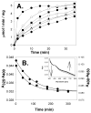
The [Fe-S] cluster associated with ApbC rapidly changes form under anoxic conditions in the absence of DTT. Panel A: ApbC-dependent Leu1 activation 2(●), 40(○), 80(■), 120(□), 180(▲), 240()), and 300(♦) minutes after desalting of reconstituted ApbC. ApbC was reconstituted as described in the materials and methods, but DTT was not added back to the sample after desalting. Panel B: The normalized absorbance changes at 325 nm (●, left axis) and 650 nm (■, right axis) as a function of time post desalting. The data in Panel B were fit by Equation 2. Panel B inset: The UV-visible absorption spectra of reconstituted ApbC after 2, 40, 80, 120, 180, 240, and 300 minutes post desalting.

References

    1. Lill R, Muhlenhoff U. Iron-sulfur protein biogenesis in eukaryotes: components and mechanisms. Annu Rev Cell Dev Biol. 2006;22:457–486. - PubMed
    1. Beinert H. Iron-sulfur proteins: ancient structures, still full of surprises. Journal of Biological and Inorganic Chemistry. 2000;5:2–15. - PubMed
    1. Frazzon J, Dean DR. Formation of iron-sulfur clusters in bacteria: an emerging field in bioinorganic chemistry. Current Opinion in Chemical Biology. 2003;7:166–173. - PubMed
    1. Takahashi Y, Tokumoto U. A third bacterial system for the assembly of iron-sulfur clusters with homologs in archaea and plastids. J Biol Chem. 2002;277:28380–28393. - PubMed
    1. Zheng M, Wang X, Templeton LJ, Smulski DR, LaRossa RA, Storz G. DNA microarray-mediated transcriptional profiling of the Escherichia coli response to hydrogen peroxide. J Bacteriol. 2001;183:4562–4570. - PMC - PubMed

Publication types

MeSH terms

LinkOut - more resources