Review
. 2008:24:183-209.
doi: 10.1146/annurev.cellbio.24.110707.175235.

Communication between the synapse and the nucleus in neuronal development, plasticity, and disease

Affiliations
Review

Communication between the synapse and the nucleus in neuronal development, plasticity, and disease

Sonia Cohen et al. Annu Rev Cell Dev Biol. 2008.

Abstract

Sensory experience is critical for the proper development and plasticity of the brain throughout life. Successful adaptation to the environment is necessary for the survival of an organism, and this process requires the translation of specific sensory stimuli into changes in the structure and function of relevant neural circuits. Sensory-evoked activity drives synaptic input onto neurons within these behavioral circuits, initiating membrane depolarization and calcium influx into the cytoplasm. Calcium signaling triggers the molecular mechanisms underlying neuronal adaptation, including the activity-dependent transcriptional programs that drive the synthesis of the effector molecules required for long-term changes in neuronal function. Insight into the signaling pathways between the synapse and the nucleus that translate specific stimuli into altered patterns of connectivity within a circuit provides clues as to how activity-dependent programs of gene expression are coordinated and how disruptions in this process may contribute to disorders of cognitive function.


Conflict of interest statement

Disclosure Statement: The authors are not aware of any biases that might be perceived as affecting the objectivity of this review.

Figures

Figure 1
Figure 1
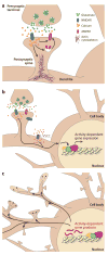
Bidirectional communication between the synapse and the nucleus mediates neuronal development and plasticity. Calcium influx into the postsynaptic cell in response to sensory experience modulates neuronal function both by direct actions at the activated synapse and through communication to the nucleus to affect activity-dependent transcriptional programs. (a) Synaptic activity induces glutamate release into the synaptic cleft and activation of the postsynaptic NMDA receptor (NMDAR). Calcium influx into the dendritic spine through the NMDA receptor regulates dendritic patterning and synapse morphology through local effects on the actin cytoskeleton. NMDA receptor activation also regulates the recruitment of AMPA receptors to the synapse in processes important for synaptic maturation and plasticity. (b) Synaptic activity is communicated to the nucleus to regulate activity-dependent gene expression. Calcium influx through both NMDA receptors and L-type voltage-sensitive calcium channels (L-VSCCs) acts as a second messenger in the cytoplasm to initiate signaling to the nucleus, where the modulation of transcription factors results in activity-dependent changes in gene expression. (c) The mRNA and protein products of activity-dependent genes regulate a range of neuronal functions in response to extracellular stimuli. During processes important for neuronal development and plasticity, the activity of these gene products throughout the cell provides a mechanism by which the nucleus is able to communicate to the synapse the functional changes required for adaptive response.
Figure 2
Figure 2
Mechanisms to increase calcium levels in the postsynaptic cell. Calcium plays a well-defined role in the biochemical transduction of signals from the synapse to the nucleus. In response to synaptic activity and neurotransmitter release, extracellular calcium flows into the postsynaptic cell through synaptic and extrasynaptic ligand- and voltage-gated calcium channels. Major routes of entry with well-established effects on nuclear gene expression are the NMDA receptor (NMDAR) and the L-type voltage-sensitive calcium channel (L-VSCC). Calcium-permeable AMPA receptors (AMPAR) may play a role at developing synapses or after the induction of synaptic plasticity. Calcium signals can also be amplified by calcium-induced release of calcium from intracellular stores, triggered by activation of ryanodine receptors (RyR). Calcium at the mouth of the channel, in the cytoplasm, or within the nucleus can signal to activity-dependent transcription factors. Alterations in calcium influx into the postsynaptic cell during development or as a result of mutation modulate the induction of gene expression in response to neuronal activity. ER denotes endoplasmic reticulum.
Figure 3
Figure 3
Model of calcium-dependent phosphorylation of CREB (cAMP response element binding protein). Phosphorylation of CREB at serine-133 in response to a diverse array of extracellular stimuli results in CREB transcriptional activation. The signal transduction cascades initiated by these stimuli ultimately result in the activation of a CREB kinase, including protein kinase A (PKA), calcium/calmodulin-dependent kinases II and IV (CaMKII and CaMKIV), and Ras/ERK-dependent kinases such as RSK. Activation of CREB-dependent transcription at particular target genes depends on additional events including other sites of CREB phosphorylation and the recruitment of transcriptional cofactors. CaMKK, CaMK kinase.
Figure 4
Figure 4
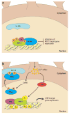
Model of calcium-dependent regulation of myocyte enhancer factor 2 (MEF2) transcriptional activity. MEF2 proteins bound to their target genes can act as either transcriptional activators or repressors, depending on the stimulation state of the cell. (a) In the unstimulated cell, the class II histone deacetylases (HDACs), which repress transcription by removing acetyl groups from histones and transcription factors, associate with MEF2. Under these conditions, MEF2 is also phosphorylated at a number of sites. Both basal phosphorylation of MEF2 at serine-408 and its association with HDACs contribute to MEF2 transcriptional repression, in part by promoting the sumoylation (Su) of MEF2 at lysine-403, a modification that represses MEF2-dependent transcription. (b) In response to synaptic activity, two calcium-dependent signaling pathways convert MEF2 from a repressor to an activator of transcription. Calcium/calmodulin-dependent protein kinase (CaMK) activation leads to the phosphorylation of the class II HDACs, initiating their binding to the 14-3-3 chaperone proteins and subsequent nuclear export. As a result, MEF2 is able to interact with the transcription-activating histone acetyltransferases (HATs), which likely increases histone acetylation at MEF2 target genes, promoting transcription, and may also contribute to the acetylation of MEF2 itself. In addition, activation of calcineurin (CaN), a calcium-dependent protein phosphatase, dephosphorylates MEF2 at serine-408. Serine-408 dephosphorylation of MEF2 promotes the desumoylation and subsequent acetylation of MEF2 lysine-403, contributing to MEF2 transcriptional activation.
Figure 5
Figure 5
Model of calcium-dependent nuclear factor of activated T cells (NFAT) activation. In the unstimulated cell, NFAT transcription factors are maintained in the cytoplasm by kinases that phosphorylate a number of NFAT phosphorylation sites. Calcineurin (CaN) docking and the subsequent dephosphorylation of NFAT induce a conformational change in the transcription factor that exposes its nuclear localization signal (NLS) and leads to NFAT transport into the nucleus. Within the nucleus, NFAT is subject to regulation by kinases that promote the export of NFAT back into the cytoplasm, thereby resulting in the shutoff of NFAT target genes. The exportin protein Crm1 shuttles NFAT back into the cytoplasm and interacts with the same region of NFAT as does calcineurin, competing with calcineurin for binding to NFAT. Nuclear NFAT kinases that phosphorylate NFAT, such as glycogen synthase kinase-3 (GSK-3), may trigger the release of calcineurin from NFAT to promote NFAT export from the nucleus. When overexpressed in Down syndrome as the result of duplication of the Down syndrome critical region (DSCR), both DSCR1 and DYRK1A [dual-specificity tyrosine (Y) phosphorylation–regulated kinase 1A] are predicted to prevent NFAT activation in response to calcium. Increased DSCR1 activity may block calcineurin-dependent NFAT dephosphorylation and translocation to the nucleus, whereas overexpression of DYRK1A may promote premature GSK3-dependent export of NFAT from the nucleus, inhibiting NFAT transcriptional activity.
Figure 6
Figure 6
Human diseases of cognitive function disrupt communication between the synapse and the nucleus. The signaling mechanisms that operate within neurons to relay the effect of synaptic stimulation to the nucleus, and the gene products produced as a result, allow communication between the synapse and the nucleus. Mutations in components of these activity-dependent signaling networks have been identified and shown to disrupt experience-dependent neuronal development and plasticity. These include mutations in the L-VSCC in Timothy syndrome, RSK2 in Coffin-Lowry syndrome, CBP in Rubenstein-Taybi syndrome, and MeCP2 in Rett syndrome. Duplications in DSCR1 and DYRK1A may contribute to the etiology of Down syndrome. Disruptions of the activity-dependent genes and their products may contribute to disorders of adaptive behavior, including depression, anxiety, and addiction. These and other mutations suggest that further insight into the programs of activity-dependent gene expression and their regulation may aid in our understanding of CNS development and function as well as of human disorders of cognitive function. Abbreviations used: CaN, calcineurin; CREB, cAMP response element binding protein; CBP, CREB-binding protein; DSCR1, Down syndrome critical region 1; DYRK1A, dual-specificity tyrosine (Y) phosphorylation–regulated kinase 1A; GSK-3, glycogen synthase kinase-3; L-VSCC, L-type voltage-sensitive calcium channel; NFAT, nuclear factor of activated T cells; MeCP2, methyl-CpG-binding protein 2; MEF2, myocyte enhancer factor 2; RSK2, ribosomal S6 kinase 2.

References

    1. Aid T, Kazantseva A, Piirsoo M, Palm K, Timmusk T. Mouse and rat BDNF gene structure and expression revisited. J Neurosci Res. 2007;85:525–35. - PMC - PubMed
    1. Aizawa H, Hu SC, Bobb K, Balakrishnan K, Ince G, et al. Dendrite development regulated by CREST, a calcium-regulated transcriptional activator. Science. 2004;303:197–202. - PubMed
    1. Alvarez VA, Sabatini BL. Anatomical and physiological plasticity of dendritic spines. Annu Rev Neurosci. 2007;30:79–97. - PubMed
    1. Arron JR, Winslow MM, Polleri A, Chang CP, Wu H, et al. NFAT dysregulation by increased dosage of DSCR1 and DYRK1A on chromosome 21. Nature. 2006;441:595–600. - PubMed
    1. Bading H, Ginty DD, Greenberg ME. Regulation of gene expression in hippocampal neurons by distinct calcium signaling pathways. Science. 1993;260:181–86. - PubMed

Publication types

MeSH terms

LinkOut - more resources