. 2008 Jul 10;1(1):21.
doi: 10.1186/1756-3305-1-21.

A monoclonal antibody marker for the exclusion-zone filaments of Trypanosoma brucei

Affiliations

A monoclonal antibody marker for the exclusion-zone filaments of Trypanosoma brucei

Mélanie Bonhivers et al. Parasit Vectors. .

Abstract

Background: Trypanosoma brucei is a haemoflagellate pathogen of man, wild animals and domesticated livestock in central and southern Africa. In all life cycle stages this parasite has a single mitochondrion that contains a uniquely organised genome that is condensed into a flat disk-like structure called the kinetoplast. The kinetoplast is essential for insect form procyclic cells and therefore is a potential drug target. The kinetoplast is unique in nature because it consists of novel structural proteins and thousands of circular, interlocking, DNA molecules (kDNA). Secondly, kDNA replication is critically timed to coincide with nuclear S phase and new flagellum biogenesis. Thirdly, the kinetoplast is physically attached to the flagellum basal bodies via a structure called the tripartite attachment complex (TAC). The TAC consists of unilateral filaments (within the mitochondrion matrix), differentiated mitochondrial membranes and exclusion-zone filaments that extend from the distal end of the basal bodies. To date only one protein, p166, has been identified to be a component of the TAC.

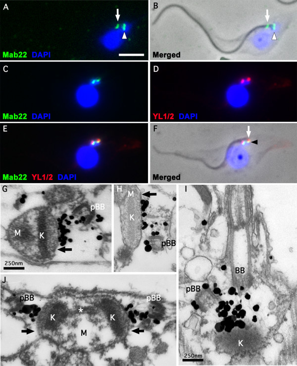
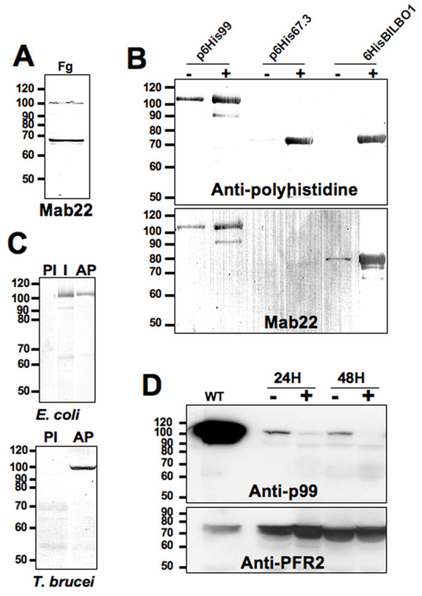
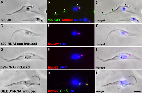
Results: In the work presented here we provide data based on a novel EM technique developed to label and characterise cytoskeleton structures in permeabilised cells without extraction of mitochondrion membranes. We use this protocol to provide data on a new monoclonal antibody reagent (Mab 22) and illustrate the precise localisation of basal body-mitochondrial linker proteins. Mab 22 binds to these linker proteins (exclusion-zone filaments) and provides a new tool for the characterisation of cytoskeleton mediated kinetoplast segregation.

Conclusion: The antigen(s) recognised by Mab 22 are cytoskeletal, insensitive to extraction by high concentrations of non-ionic detergent, extend from the proximal region of basal bodies and bind to the outer mitochondrial membrane. This protein(s) is the first component of the TAC exclusion-zone fibres to be identified. Mab 22 will therefore be important in characterising TAC biogenesis.


Figures

Figure 1
Figure 1
Immunofluorescence and Immuno-electron microscope labelling of the T. brucei TAC. A-F. Immunofluorescence on wild-type cytoskeletons. A. DAPI staining (blue) and Mab 22 labelling (green). B. A merged phase contrast, DAPI staining and Mab 22 labelling of the cytoskeleton in A. The arrows in A and B show the Mab 22 flagellar pocket collar label observed when this monoclonal was initially made and presumably had a very high titre. The arrowhead of A and B indicate the TAC exclusion zone label of Mab 22. C-D. A double labelling immunofluorescence micrograph illustrating a cytoskeleton showing Mab 22 and YL1/2 label. C. DAPI staining (blue) and Mab 22 labelling (green). D. DAPI staining (blue) and anti-basal body staining using YL1/2 (red). E. Merged of C and D. F. Phase contrast merged of E. The arrow in F illustrates the basal body labelling of YL1/2 and the arrowhead indicates the TAC exclusion zone label. In A-F scale bar is 5 μm. G-J. Electron micrographs of longitudinal sections of extracted and Mab 22 probed cytoskeletons. In all images the section traverse a basal and/or pro-basal bodies (BB, pBB). The immunolabelling is clearly observed between the basal or probasal bodies and the outer mitochondrion membrane (black arrow). Kinetoplast is marked as (K) and mitochondrion matrix marked as (M). In J, the kinetoplast is in early kinetoplast S phase and illustrates the presence of unilateral filaments and attachment to the mitochondrion membrane and kinetoplast. The asterisk in J denotes the "nabelschnur", a filamentous structure rich in basic proteins that links the kDNA discs during their segregation [18]. In G-J scale bar is 250 nm.
Figure 2
Figure 2
Western blots. A. Flagellar proteins (5.108 flagella, Fg) probed with Mab 22. B. N-terminal Histidine-tagged proteins expressed in E. coli (2.107 bacteria/well) and probed with anti-polyhistidine (upper panel) and Mab 22 (lower panel). Non-induced bacteria (-), 3 hr IPTG induction (+). C. Specificity of the affinity purified polyclonal anti-p99. In upper and lower panels PI: Pre-immune serum 1:1,000. I: Immune serum 1:500, AP: affinity purified immune serum 1:1000. WC: T. brucei whole cells (107/well). D. p99 RNAi whole cells (4.107/well) probed with the affinity purified immune serum anti-p99 1:500 and anti-PFR2 L8C4 monoclonal antibody (1:1,000) as loading control. Non-induced cells (-), Induced cells (+), Wild-type cells (WT).
Figure 3
Figure 3
GFP tagged and immunofluorescence on RNAi knockdown cells. A-C. 48 h induced p99-GFP expressing cytoskeletons (green) probed with Mab 22 (red) and DAPI (Blue). We made a GFP tagged protein because the rabbit antibody raised to the 99 kDa was positive by Western blots on E. coli expressed purified proteins and trypanosome cells but was not detectible by immunofluorescence. D-F. Non-induced p99 RNAi cytoskeletons probed with Mab 22 and DAPI (Blue). G-I. 48 h induced p99 RNAi cytoskeletons probed by Mab 22 and DAPI (Blue). K-L. BILBO1 24 h induced RNAi cytoskeletons probed with Mab 22 and YL1/2 and DAPI (Blue). Note that the Mab 22 antibody signal remains present after probing p99 and BILBO1 RNAi induced knockdown cytoskeletons indicating that the gene expressing the TAC protein is neither p99 nor BILBO1. In A-L scale bar is 5 μm.

References

    1. Sherwin T, Schneider A, Sasse R, Seebeck T, Gull K. Distinct localization and cell cycle dependence of COOH terminally tyrosinolated alpha-tubulin in the microtubules of Trypanosoma brucei brucei. J Cell Biol. 1987;104:439–446. doi: 10.1083/jcb.104.3.439. - DOI - PMC - PubMed
    1. Sherwin T, Gull K. The cell division cycle of Trypanosoma brucei brucei: timing of event markers and cytoskeletal modulations. Philos Trans R Soc Lond B Biol Sci. 1989;323:573–588. doi: 10.1098/rstb.1989.0037. - DOI - PubMed
    1. Robinson DR, Sherwin T, Ploubidou A, Byard EH, Gull K. Microtubule polarity and dynamics in the control of organelle positioning, segregation, and cytokinesis in the trypanosome cell cycle. J Cell Biol. 1995;128:1163–1172. doi: 10.1083/jcb.128.6.1163. - DOI - PMC - PubMed
    1. Robinson DR, Gull K. Basal body movements as a mechanism for mitochondrial genome segregation in the trypanosome cell cycle. Nature. 1991;352:731–733. doi: 10.1038/352731a0. - DOI - PubMed
    1. Kohl L, Robinson D, Bastin P. Novel roles for the flagellum in cell morphogenesis and cytokinesis of trypanosomes. Embo J. 2003;22:5336–5346. doi: 10.1093/emboj/cdg518. - DOI - PMC - PubMed

LinkOut - more resources