

Review
doi: 10.1016/j.drudis.2008.06.006.
Epub 2008 Aug 27.
Limitations and lessons in the use of X-ray structural information in drug design
Affiliations
- PMID: 18617015
- PMCID: PMC7185550
- DOI: 10.1016/j.drudis.2008.06.006
Item in Clipboard
Review
Limitations and lessons in the use of X-ray structural information in drug design
Drug Discov Today.
2008 Oct.
Abstract
The use of X-ray crystal structure models continues to provide a strong stimulus to drug discovery, through the direct visualisation of ligand–receptor interactions. There is sometimes a limited appreciation of the uncertainties introduced during the process of deriving an atomic model from the experimentally observed electron density. Here, some of these uncertainties are highlighted with recent examples from the literature, together with snippets of advice for the medicinal chemist embarking on using X-ray crystal structure information in a drug discovery programme.
Figures
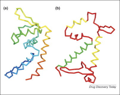
(a) Cα-trace, colour-ramped from blue at the N-terminus to red at the C-terminus, of the correct structure of the SarA protein (wwPDB code 2FRH). (b) Cα-trace, shown in the same orientation as (a), of an incorrect model of the same protein determined six years earlier (wwPDB code 1FZN). The N-terminal helix has been coloured green as it has the correct secondary structure and the correct sequence registration. The two yellow helices have counterparts in the correct structure but their sequence assignment is incorrect.
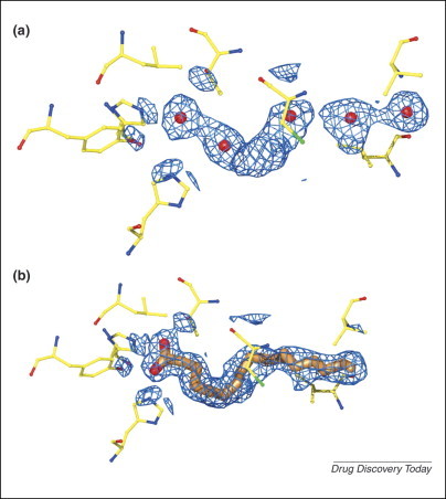
(a) Electron density in the active site of what was assumed to be an apo-form of PPAR-β/δ was modelled by several water molecules (red spheres) that led to the puzzling conclusion that the apo-structure displayed the conformation of the activated (ligand-bound) state of the protein (wwPDB code 2GWX). (b) A re-evaluation of the experimental data of the original study led to the identification of a bound fatty acid ligand (cis-vaccenic acid) that suddenly explained the earlier conundrum (wwPDB code 2BAW). The ligand is shown with gold carbon atoms. All residues that have at least one atom within 3.5A of any ligand atom have been included (the same residues were also included in (a)). The electron-density map (retrieved from EDS) is shown within 2.0 Å of any ligand atom. The EDS density in (a) is shown within 2.0 Å of any ligand atom if it had been modelled as in (b).
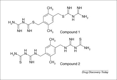
Compound 1 the actual inhibitor, and compound 2 the putative inhibitor of dihydrofolate reductase identified by HTS.
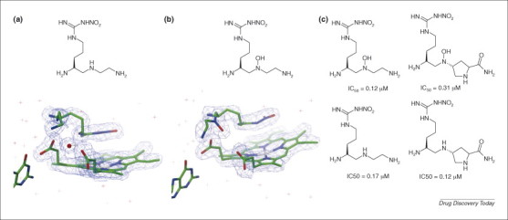
(a) Crystallographic data showed the active site of nNOS contained a substituted arginine mimic bound over the haem, and electron density consistent with a water molecule that potentially could be displaced. (wwPDB code 2HX3) Structure model shown with 2Fo-Fc map contoured at 1σ⋅ (b) The design and synthesis of an N-hydroxy arginine mimic proved successful, and crystallographic data confirmed that the water molecule had indeed been replaced (wwPDB code 1P6I) Structure model shown with 2Fo-Fc map contoured at 1σ. (c) Contrary to expectations, structure–activity relationships showed little benefit in displacing the active-site water molecule.
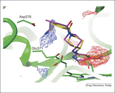
Mouse iNOS protein (green) complexed with the GOLD docking of the designed compound (yellow) and the X-ray structure of this compound (purple). The GRID density for a N3+ probe (cationic nitrogen) at contour level −12 with LEAU = 0 is shown as a blue grid. The red grid shows where the N3+ probe is unfavourable compared with a water molecule at contour level +1 with LEAU = 3. Note that the red density is not in the region of the amine designed to interact with Asp376.
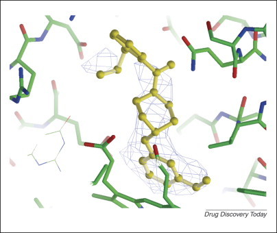
The structure model of the designed compound shown with the 2Fo-Fc map contoured at 1σ, showing only weak density around the putative position of the ethylamine sidechain.
References
-
- Rowland P. Crystal structure of human cytochrome P450 2D6. J. Biol. Chem. 2006;281:7614–7622. - PubMed
-
- Yano J.K. The structure of human microsomal cytochrome P450 3A4 determined by X-ray crystallography to 2.05-A resolution. J. Biol. Chem. 2004;279:38091–38094. - PubMed
-
- Williams P.A. Crystal structures of human cytochrome P450 3A4 bound to metyrapone and progesterone. Science. 2004;305:683–686. - PubMed
-
- Williams P.A. Crystal structure of human cytochrome P450 2C9 with bound warfarin. Nature. 2003;424:464–468. - PubMed
Publication types
MeSH terms
Substances
LinkOut - more resources
Full Text Sources
