. 2008 Sep 5;283(36):24497-505.
doi: 10.1074/jbc.M802825200. Epub 2008 Jul 10.

Phosphorylation of Thr-178 and Thr-184 in the TAK1 T-loop is required for interleukin (IL)-1-mediated optimal NFkappaB and AP-1 activation as well as IL-6 gene expression

Affiliations

Phosphorylation of Thr-178 and Thr-184 in the TAK1 T-loop is required for interleukin (IL)-1-mediated optimal NFkappaB and AP-1 activation as well as IL-6 gene expression

Yang Yu et al. J Biol Chem. .

Abstract

TAK1 (transforming growth factor-beta-activated kinase 1), a mitogen-activated protein kinase kinase kinase, is activated by various cytokines, including interleukin-1 (IL-1). However, the precise regulation for TAK1 activation at the molecular level is still not fully understood. Here we report that dual phosphorylation of Thr-178 and Thr-184 residues within the kinase activation loop of TAK1 is essential for TAK1-mediated NFkappaB and AP-1 activation. Once co-overexpressed with TAB1, TAK1 mutant with alanine substitution of these two residues fails to activate IKKbeta-mediated NFkappaB and JNK-mediated AP-1, whereas TAK1 mutant with replacement of these two sites with acidic residues acts like the TAK1 wild type. Consistently, TAK1 mutant with alanine substitution of these two residues severely inhibits IL-1-induced NFkappaB and AP-1 activities, whereas TAK1 mutant with replacement of these two sites with acidic residues slightly enhances IL-1-induced NFkappaB and AP-1 activities compared with the TAK1 wild-type. IL-1 induces the phosphorylation of endogenous TAK1 at Thr-178 and Thr-184. Reconstitution of TAK1-deficient mouse embryo fibroblast cells with wild-type TAK1 or a TAK1 mutant containing threonine 178 and 184 to alanine mutations revealed the importance of these two sites in IL-1-mediated IKK-NFkappaB and JNK-AP-1 activation as well as IL-1-induced IL-6 gene expression. Our finding is the first report that substitution of key serine/threonine residues with acidic residues mimics the phosphorylated state of TAK1 and renders TAK1 active during its induced activation.


Figures
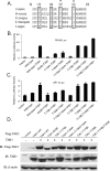
FIGURE 1.
FIGURE 1.
Thr-178 and Thr-184 residues within the activation loop of human TAK1 are potential phosphorylation sites required for the TAK1/TAB1-induced NFκB and AP-1 activation. A, sequence alignment of kinase activation loop of TAK1 from different species. The potential phosphorylation sites are marked with an open box. Thr-187 and Ser-192 have been reported previously and are marked with a star. B and C, the effect of alanine or glutamic acid substitution at the indicated residue within the activation loop on the TAK1-induced NFκB(B) and AP-1 (C) activities. One microgram of NFκBor AP-1 luciferase reporter plasmid and 20 ng of Renilla luciferase plasmid were cotransfected into TAK1-deficient cells with 1 μg of empty vector control or different TAK1 expression plasmids. The relative luciferase activity was measured 36 h later and normalized with the Renilla activity. Error bars, ±S.D. in triplicate experiments. D, expression levels of FLAG-TAK1 and TAB1 in the transfected cells were detected by immunoblotting, respectively. β-Actin was detected as a loading control.
FIGURE 2.
FIGURE 2.
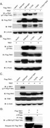
Generation and characterization of an antibody specific for phospho-TAK1 at Thr-178 and Thr-184. A, the anti-phospho-TAK1 (pT178/pT184) antibody is specific for human TAK1 phospho-Thr-178/Thr-184. Cell extracts were prepared from HEK293T cells transfected with expression vectors for TAB1 as well as the FLAG-TAK1 and its derived mutants as indicated and analyzed by immunoblotting with the anti-phospho-TAK1 antibody (pT178/pT184) (top) and reprobed with an anti-FLAG and anti-TAB1 antibodies to detect the expression level of FLAG-TAK1 and TAB1, respectively. β-Actin was detected as a loading control (bottom). B, the anti-phospho-TAK1 (pT184) antibody detects the phosphorylated form of human TAK1 at Thr-184. Cell extracts were prepared from HEK293T cells transfected with an expression vector for TAB1 as well as the FLAG-TAK1 and its derived mutants as indicated and analyzed by immunoblotting with the anti-phospho-TAK1 antibody (pT184) and reprobed with anti-FLAG and anti-TAB1 antibodies to detect the expression level of FLAG-TAK1 and TAB1, respectively. β-Actin was detected as a loading control. C, Ala substitution at Thr-187 and Ser-192 abolished TAK1/TAB1 overexpression-induced TAK1 phosphorylation at Thr-178 and Thr-184. Cell extracts were prepared from HEK293T cells transfected with an expression vector for TAB1 as well as the FLAG-TAK1, as indicated, and analyzed by immunoblotting with the anti-phospho-TAK1 antibody (pT178/pT184) and reprobed with anti-FLAG and anti-TAB1 antibodies to detect the expression level of FLAG-TAK1 and TAB1, respectively. β-Actin was detected as a loading control. D, the anti-phospho-TAK1(pT178/pT184) antibody detects the phosphorylated form of human TAK1 at Thr-178 and Thr-184. Cell extracts were prepared from HEK293T cells transfected with an expression vector for FLAG-TAK1 and TAB1 and were immunoprecipitated with anti-FLAG antibody and then treated with or without λ-protein phosphatase (λ-PPase) for 30 min before being analyzed by immunoblotting with the anti-phospho-TAK1 antibody and reprobed with anti-FLAG antibody.
FIGURE 3.
FIGURE 3.
Phosphorylation of TAK1 at Thr-178 and Thr-184 residues is required for the TAK1-mediated IKK-NFκB and JNK-AP-1 activations. A, phosphorylation of both Thr-178 and Thr-184 residues is essential for TAK1-mediated phosphorylation of IKKβ. TAK1-deficient (–/–) MEF cells were cotransfected with empty vector or FLAG-TAK1 wild-type expression vector or derived mutants in the presence of HA-IKKβ and TAB1 using Lipofectamine 2000. After 36 h, cell extracts were immunoprecipitated (IP) with anti-HA and immunoblotted (IB) with anti-phospho-IKKα/β (p-IKKβ). HA-IKKβ proteins present in the immunoprecipitates were detected by stripping the blots and reprobing with anti-HA antibody. The expression level of FLAG-TAK1 wild-type and derived mutated proteins were detected by anti-FLAG antibody. B, HEK293T cells were transiently transfected with empty vector, TAK1 wild type, and the indicated mutants. Cells were collected 36 h after transfection and lysed with protein lysis buffer, and the same amount of protein was subjected to SDS-PAGE followed by immunoblotting analysis to detect the phospho-JNK and phospho-p38 proteins in the cell lysates using antibodies specific for the phosphorylated form of JNK and p38, respectively. The same membranes were stripped and reprobed with anti-JNK and anti-p38 antibodies to detect the level of total JNK and p38 expression. The expression of TAK1 was analyzed by immunoblotting with the antibodies indicated. β-Actin was detected as a loading control. C and D, phosphorylation of both Thr-178 and Thr-184 residues is required for TAK1-induced NFκB(C) and AP-1 (D) luciferase activities in the TAK1-deficient MEF cells. One microgram of NFκB or AP-1 luciferase reporter plasmid and 20 ng of Renilla luciferase plasmid as well as TAB1 expression plasmid were transfected into the deficient MEF cells, along with 1 μg of empty vector or expression plasmid encoding the wild type or the indicated TAK1 mutants. The relative luciferase activity was measured 36 h later and normalized with the Renilla activity. Error bars, ±S.D. in triplicate experiments.
FIGURE 4.
FIGURE 4.
Phosphorylation of TAK1 at both Thr-178 and Thr-184 residues is required for IL-1-mediated IKK-NFκB and JNK-AP-1 activations. A, IL-1 induces the phosphorylation of TAK1 at Thr-178 and Thr-184 residues. HeLa cells were transfected with the expression plasmid encoding TAK1. 48 h after transfection, cells were either untreated or treated with IL-1 (10 ng/ml) before being harvested and lysed. FLAG-tagged TAK1 protein in the cell lysates was immunoprecipitated (IP) with antibody specific for FLAG tag and subsequently subjected to immunoblotting analysis (IB) with the anti-phospho-TAK1 (pT178/pT184) antibody to detect the level of phosphorylated TAK1 (top). The same blot was stripped and reprobed with an anti-FLAG antibody to detect the level of immunoprecipitated total TAK1 protein (bottom). B and C, expression of the TAK1 T178A/T184A mutant inhibits IL-1-induced NFκB and AP-1 reporter activities. One μg of NFκB (NFκB-Luc) or AP-1 (AP-1-Luc) reporter and 20 ng of Renilla-Luc plasmid were cotransfected into TAK1-deficient MEF cells with control vector or TAK1 wild-type expression vector or derived mutants for 24 h followed by the addition of IL-1 (10 ng/ml) for 12 h. Cell extracts were collected to determine the relative luciferase activity. Error bars, ±S.D. in triplicate experiments. D, expression of the TAK1 T178A/T184A mutant inhibits the IL-1-induced JNK, p38, IKK, IκBα, and NFκB-p65 phosphorylation as well as IκBα degradation and NFκB nuclear translocation. TAK1-deficient MEF cells were transduced with the retrovirus encoding the vector control, TAK1 wild type, or TAK1 T178A/T184A mutant and subsequently selected with puromycin (2 μg/ml) to establish the TAK1-deficient MEF cell lines with the stable expression of either TAK1 wild type or T178A/T184A mutant. TAK1-deficient, reconstituted wild-type and T178A/T184A MEF cells were untreated or treated with IL-1 (10 ng/ml) for the time points indicated and subsequently harvested. Whole cell extracts and nuclear extracts were subjected to SDS-PAGE and immunoblotted with the antibodies indicated. β-Actin was detected as a loading control for whole cell extracts, and proliferating cell nuclear antigen was used as a loading control for nuclear extracts. E and F, expression of the TAK1 T178A/T184A mutant inhibits IL-1-induced NFκB and AP-1 reporter activities. One μg of NFκB (E) or AP-1 (F) reporter and 20 ng of Renilla-Luc plasmid were cotransfected into TAK1-deficient MEF stable cell lines with control or TAK1 wild-type or T178A/T184A mutant expression vectors for 24 h followed by the addition of IL-1 (10 ng/ml) for 12 h. The relative luciferase activity was measured and normalized with the Renilla activity. Error bars, ±S.D. in triplicate experiments.
FIGURE 5.
FIGURE 5.
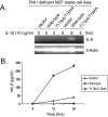
Phosphorylation of TAK1 at Thr-178 and Thr-184 residues is required for IL-1-induced IL-6 gene expression. A, expression of the TAK1 T178A/T184A mutant inhibits IL-1-induced IL-6 gene transcription. TAK1-deficient, wild-type, and T178A/T184A MEF cells were untreated or treated with IL-1 (10 ng/ml) for 8 h and subsequently harvested for extraction of total RNA using Trizol reagent. One μg of total RNA was used to synthesize first-strand cDNA using a reverse transcription kit according to the manufacturer's instructions. These synthesized cDNAs were used as templates for mouse IL-6 PCR amplification. The PCR products were resolved in 2% agarose gel. B, expression of the TAK1 T178A/T184A mutant inhibits IL-1-induced IL-6 production. TAK1-deficient, wild-type, and T178A/T184A MEF cells were untreated or treated with IL-1 (10 ng/ml) for the times indicated. The supernatants from these cultures were collected and subjected to mouse IL-6 enzyme-linked immunosorbent assay analysis according to the manufacturer's instructions.
FIGURE 6.
FIGURE 6.
A working model for TAK1 action in the IL-1-mediated IKK-NFκB and JNK-AP-1 activation. IL-1β induces TAK1 phosphorylation at four sites within the kinase activation loop and subsequent activation in the cells to mediate optimal IKK-NFκB and JNK-AP-1 activation as well as NFκB- and AP-1-dependent IL-6 gene expression.

Similar articles

Cited by

References

    1. Dinarello, C. A. (1997) Cytokine Growth Factor Rev. 8 253–265 - PubMed
    1. O'Neill, L. A., and Greene, C. (1998) J. Leukocyte Biol. 63 650–657 - PubMed
    1. Arend, W. P. (2002) Cytokine Growth Factor Rev. 13 323–340 - PubMed
    1. Beutler, B. (2004) Nature 430 257–263 - PubMed
    1. Baud, V., and Karin, M. (2001) Trends Cell Biol. 11 372–377 - PubMed

Publication types

MeSH terms