Comparative Study
. 2008 Sep;295(3):G470-8.
doi: 10.1152/ajpgi.90262.2008. Epub 2008 Jul 10.

Differences in activity and phosphorylation of MAPK enzymes in esophageal squamous cells of GERD patients with and without Barrett's esophagus

Affiliations
Comparative Study

Differences in activity and phosphorylation of MAPK enzymes in esophageal squamous cells of GERD patients with and without Barrett's esophagus

Hui Ying Zhang et al. Am J Physiol Gastrointest Liver Physiol. 2008 Sep.

Abstract

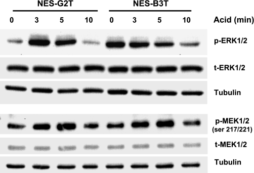
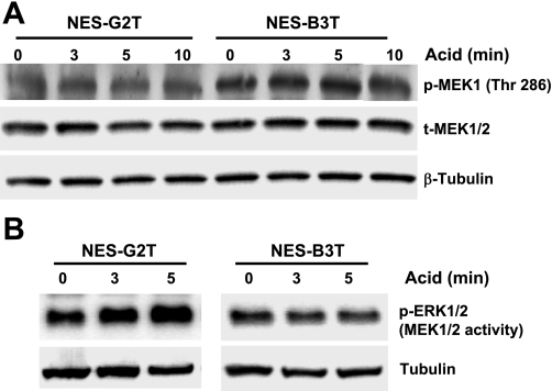
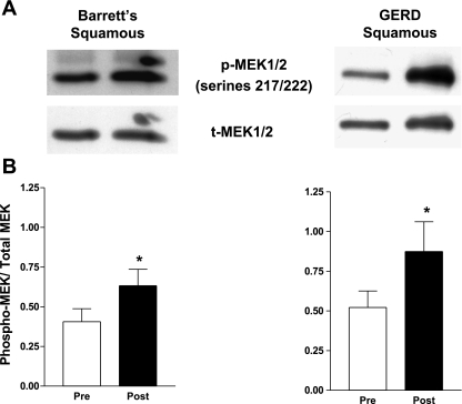
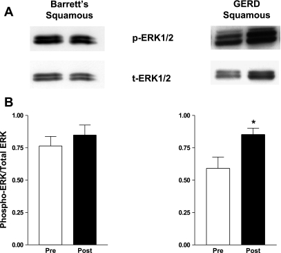
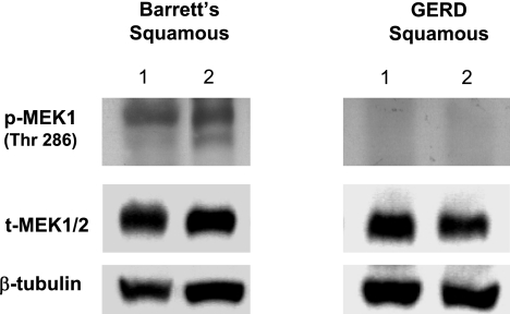
We hypothesized that, in esophageal squamous epithelial cells, there are differences among individuals in the signal transduction pathways activated by acid reflux that might underlie the development of Barrett's esophagus. To explore that hypothesis, we immortalized nonneoplastic, esophageal squamous cells from patients with gastroesophageal reflux disease (GERD) with (NES-B3T) and without (NES-G2T) Barrett's esophagus and used those cells to study acid effects on MAPK proteins. During endoscopy in patients with GERD with and without Barrett's esophagus, we took biopsy specimens from the distal squamous esophagus to study MAPK proteins before and after esophageal perfusion with 0.1 N HCl. We used immunoblotting and Western blotting to study MEK1/2 phosphorylation at two activating sites (serines 217/221), MEK1 phosphorylation at an inhibitory site (threonine 286), and MEK1/2 activity. After acid exposure, both cell lines exhibited increased MEK1/2 phosphorylation at the activating sites; the NES-B3T cells had higher levels of MEK1 phosphorylation at the inhibitory site, however, and only the NES-G2T cells showed an acid-induced increase in MEK1/2 activity. Similarly, in the squamous epithelium of patients with GERD with and without Barrett's esophagus, acid perfusion increased MEK1/2 phosphorylation at the activating sites in both patient groups; the Barrett's patients had higher levels of MEK1 phosphorylation at the inhibitory site, however, and only the patients without Barrett's demonstrated an acid-induced increase in ERK1/2 phosphorylation. In esophageal squamous cell lines and biopsies from patients with GERD with and without Barrett's esophagus, we have found differences in MAPK pathways activated by acid exposure. We speculate that these differences might underlie the development of Barrett's metaplasia.


Figures

Fig. 1.
Fig. 1.
Telomerase-immortalized esophageal squamous epithelial cells in culture. Low (4×) and high (10×) power magnifications of esophageal squamous epithelial cells (white arrows) grown with ATCC Albino Swiss 3T3 fibroblast feeder cells (black arrows). A: NES-G2T squamous cells from a patient with gastroesophageal reflux disease (GERD) without Barrett's esophagus. B: NES-B3T squamous cells from a patient with GERD with Barrett's esophagus.
Fig. 2.
Fig. 2.
A: telomerase activity in parent and human telomerase reverse transcriptase (hTERT)-infected esophageal squamous cell lines. HeLa cells served as a positive (+) control; lysis buffer served as a negative (−) control. TRAP assay shows a characteristic, six-base-pair ladder in cells with telomerase activity, which include the positive control and the NES-B3 and NES-G2 cells infected with hTERT. B: both the NES-G2T and NES-B3T cell lines express the squamous cytokeratin (CK4 and CK13); β-tubulin served as a loading control. Skin derived keratinocytes (Ker) served as a positive control.
Fig. 3.
Fig. 3.
Telomerase-immortalized esophageal squamous cells exhibit contact inhibition and do not grow in soft agar. A: NES-G2T and NES-B3T cells demonstrate early log phase growth, but, with increasing confluence, the cells demonstrate a growth plateau. B: NES-G2T and NES-B3T cells show no colonies after 3 wk in soft agar in contrast to SEG-1 cancer cells, which exhibit numerous colonies.
Fig. 4.
Fig. 4.
UVB irradiation induces expression of p53 and p21 in NES-G2T and NES-B3T cells. After irradiation, both cell lines show increased p53 expression by 6 h and increased p21 expression by 24 h; β-tubulin served as a loading control. MCF-7 cells treated with 0.2 μg/ml of doxorubicin served as a positive control for p53 and p21 expression.
Fig. 5.
Fig. 5.
Acid increases MEK1/2 phosphorylation at activating sites (serines 217/221) in both cell lines, but acid increases ERK1/2 phosphorylation only in NES-G2T cells. Total ERK1/2, total MEK1/2, and β-tubulin were used as loading controls.
Fig. 6.
Fig. 6.
A: NES-B3T cells have higher levels of MEK1 phosphorylation at an inhibitory site than NES-G2T cells. Total MEK1/2 and β-tubulin were used as loading controls. B: acid exposure increases MEK1/2 activity (measured as ERK1/2 phosphorylation) in NES-G2T but not in NES-B3T cells. β-tubulin expression in whole cell lysates served as a control to confirm that equal amounts of protein were added in the immunoprecipitation reaction.
Fig. 7.
Fig. 7.
Acid perfusion induces MEK1/2 phosphorylation on serines 217/222 in esophageal squamous epithelium of patients with GERD with and without Barrett's esophagus in vivo. A: representative Western blot demonstrating MEK1/2 phosphorylation and total MEK1/2 expression before and after esophageal acid perfusion. B: MEK1/2 phosphorylation relative to total MEK1/2 increases significantly following acid perfusion in esophageal squamous epithelium of patients with GERD with and without Barrett's esophagus (*P < 0.05). The bar graphs show the mean ± SE of esophageal squamous epithelial biopsies from 7 patients with GERD with Barrett's esophagus and 5 patients with GERD without Barrett's esophagus before and after acid perfusion.
Fig. 8.
Fig. 8.
Acid perfusion induces ERK1/2 phosphorylation in esophageal squamous epithelium in patients who have GERD without Barrett's esophagus. A: representative Western blot demonstrating ERK1/2 phosphorylation before and after acid perfusion. B: ERK 1/2 phosphorylation relative to total ERK 1/2 increased significantly following acid exposure in esophageal squamous epithelium from patients who had GERD without Barrett's esophagus but not in those with Barrett's esophagus (*P < 0.05). The bar graphs show the mean ± SE of squamous epithelial biopsies from 7 patients with GERD with Barrett's esophagus and 5 patients with GERD without Barrett's esophagus before and after acid perfusion.
Fig. 9.
Fig. 9.
Phosphorylation of MEK1 on threonine 286 in esophageal squamous epithelium from 2 representative patients with GERD with Barrett's esophagus and 2 representative patients with GERD without Barrett's esophagus. Note expression of the inhibitory form of phospho-MEK1 only in the patients with Barrett's esophagus. β-tubulin served as a loading control.

References

    1. Alessi DR, Gomez N, Moorhead G, Lewis T, Keyse SM, Cohen P. Inactivation of p42 MAP kinase by protein phosphatase 2A and a protein tyrosine phosphatase, but not CL100, in various cell lines. Curr Biol 5: 283–295, 1995. - PubMed
    1. Ali I, Rafiee P, Hogan WJ, Jacob HJ, Komorowski RA, Haasler GB, Shaker R. Dickkopf homologs in squamous mucosa of esophagitis patients are overexpressed compared with Barrett's patients and healthy controls. Am J Gastroenterol 101: 1437–1448, 2006. - PubMed
    1. Boch JA, Shields HM, Antonioli DA, Zwas F, Sawhney RA, Trier JS. Distribution of cytokeratin markers in Barrett's specialized columnar epithelium. Gastroenterology 112: 760–765, 1997. - PubMed
    1. Feagins LA, Zhang HY, Zhang X, Hormi-Carver K, Thomas T, Terada LS, Spechler SJ, Souza RF. Mechanisms of oxidant production in esophageal squamous cell and Barrett's cell lines. Am J Physiol Gastrointest Liver Physiol 294: G411–G417, 2008. - PubMed
    1. Glickman JN, Chen YY, Wang HH, Antonioli DA, Odze RD. Phenotypic characteristics of a distinctive multilayered epithelium suggests that it is a precursor in the development of Barrett's esophagus. Am J Surg Pathol 25: 569–578, 2001. - PubMed

Publication types

MeSH terms