. 2008 Sep;22(9):2116-27.
doi: 10.1210/me.2008-0059. Epub 2008 Jul 10.

Nuclear and extranuclear pathway inputs in the regulation of global gene expression by estrogen receptors

Affiliations

Nuclear and extranuclear pathway inputs in the regulation of global gene expression by estrogen receptors

Zeynep Madak-Erdogan et al. Mol Endocrinol. 2008 Sep.

Abstract

Whereas estrogens exert their effects by binding to nuclear estrogen receptors (ERs) and directly altering target gene transcription, they can also initiate extranuclear signaling through activation of kinase cascades. We have investigated the impact of estrogen-mediated extranuclear-initiated pathways on global gene expression by using estrogen-dendrimer conjugates (EDCs), which because of their charge and size remain outside the nucleus and can only initiate extranuclear signaling. Genome-wide cDNA microarray analysis of MCF-7 breast cancer cells identified a subset of 17beta-estradiol (E2)-regulated genes ( approximately 25%) as EDC responsive. The EDC and E2-elicited increases in gene expression were due to increases in gene transcription, as observed in nuclear run-on assays and RNA polymerase II recruitment and phosphorylation. Treatment with antiestrogen or ERalpha knockdown using small interfering RNA abolished EDC-mediated gene stimulation, whereas GPR30 knockdown or treatment with a GPR30-selective ligand was without effect, indicating ER as the mediator of these gene regulations. Inhibitors of MAPK kinase and c-Src suppressed both E2 and EDC stimulated gene expression. Of note, in chromatin immunoprecipitation assays, EDC was unable to recruit ERalpha to estrogen-responsive regions of regulated genes, whereas ERalpha recruitment by E2 was very effective. These findings suggest that other transcription factors or kinases that are downstream effectors of EDC-initiated extranuclear signaling cascades are recruited to regulatory regions of EDC-responsive genes in order to elicit gene stimulation. This study thus highlights the importance of inputs from both nuclear and extranuclear ER signaling pathways in regulating patterns of gene expression in breast cancer cells.


Figures
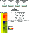
Figure 1
Figure 1
cDNA Microarray Analysis of Genes Regulated by E2 and EDC A, Experimental design. MCF-7 cells were treated with vehicle, 10 nm E2, empty dendrimer, or 10 nm E2 equivalent EDC for 4 h. RNA was then hybridized to Affymetrix Hu-U133 GeneChips, and analysis was performed as described in Materials and Methods. B, Cluster diagram of EDC- and E2-regulated genes. C, Venn diagram showing the distribution of genes regulated by E2 only, commonly regulated by E2 and EDC, or regulated by EDC only, and their characterization in functional categories according to GeneSpring and ErmineJ softwares and web-based DAVID functional annotation tool. Ctrl, Control; Dend, empty dendrimer; Veh, vehicle.
Figure 2
Figure 2
Time Course of Regulation of Gene Expression by E2 or EDC MCF-7 cells were treated with 0.1% EtOH vehicle, 10 nm E2 (solid line), empty dendrimer or 10 nm estrogen equivalent EDC (dotted line) for 2, 4, and 8 h, and mRNA levels were monitored by Q-PCR. mRNA levels were normalized relative to 36B4, and fold change was calculated relative to control. Results are the average ± sd of at least three independent experiments.
Figure 3
Figure 3
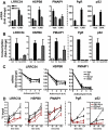
EDC and E2 Increase Gene Expression by Increasing Gene Transcription and Not Altering mRNA Stability A, MCF-7 cells were pretreated with 10 μg/ml cycloheximide for 2 h to stop protein translation and were then treated with 10 nm E2 or 10 nm E2 equivalent EDC for 4 h in the continued presence of cycloheximide. Total RNA was isolated and reverse transcribed. Q-PCR was performed. B, Nuclear run-on assays were performed on nuclei isolated from MCF-7 cells after treatment with 0.1% ethanol vehicle, 10 nm E2, empty dendrimer (Dend), or 10 nm E2 equivalent EDC for 2 h. Fold change in transcription is shown. C, MCF-7 cells were treated with 5 μg/ml actinomycin D in the presence or absence of 10 nm E2 or 10 nm E2 equivalent EDC for 2 or 4 h. Total RNA was isolated and reverse transcribed. Q-PCR was performed. D, MCF-7 cells were treated with 10 nm E2 or 10 nm E2 equivalent EDC for 15 min, 1 h, and 2 h. Total Pol II/DNA complexes or phosphor-Ser5-Pol II/DNA complexes were immunoprecipitated overnight using specific anti-Total Pol II antibody, anti-phospho-Ser5_Pol II (CTD4H8), or rabbit IgG (Santa Cruz) as negative control. Immunoprecipitated DNA levels were measured by Q-PCR, and percent input was calculated. Values are the mean ± sd of at least three independent experiments. CHX, Cycloheximide; Pol II, polymerase II; Veh, vehicle.
Figure 4
Figure 4
ERα Is the Mediator for EDC Regulation of Gene Expression A, MCF-7 cells were pretreated with 1 μm ICI182780 for 2 h and then treated with 10 nm E2 or 10 nm E2 equivalent EDC for 4 h. B, MCF-7 cells were transfected with control GL3 siRNA or ERα siRNA for 48 h and then treated with 10 nm E2 or 10 nm E2 equivalent EDC for 4 h. Western blot inset in panel B right shows the extent of ER knockdown with siRNA to be about 80%. C, MCF-7 cells were cotreated with 10 nm G1 (GPR30 agonist ligand) during 10 nm E2 or 10 nm E2 equivalent EDC treatment. D, MCF-7 cells were treated with the indicated concentrations of G1 ligand for 4 h. Total RNA was isolated, reverse transcribed, and analyzed by Q-PCR. Values are the mean ± sd of at least three independent experiments. Ctrl, Control; siERα, small interfering siERα; siGL3, control siRNA.
Figure 5
Figure 5
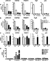
Inhibitors of c-Src Kinase or MEK Dampen E2- and EDC-Induced Gene Regulation MCF-7 cells were pretreated with 1 μm PP2 or 50 μm PD98059 for 2 h and then treated with control vehicle, 10 nm E2, empty dendrimer, or 10 nm E2 equivalent EDC for 4 h. Total RNA was isolated and reverse transcribed. Q-PCR was performed. ***, P < 0.001; or **, P < 0.01; or *, P < 0.05, significantly up-regulated by E2 or EDC over control. #, P < 0.05; or ##, P < 0.01; significantly different from no inhibitor control. Values are the mean ± sd of three or more independent experiments. Veh, Vehicle.
Figure 6
Figure 6
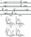
EDC Is Ineffective in Recruiting ERα to ER Binding Sites of Estrogen Target Genes whereas E2 Elicits a Robust ERα Recruitment A, Schematic showing the location of ERα binding sites for selected genes. B, MCF-7 cells were treated with 10 nm E2 or 10 nm E2 equivalent EDC for 45 min, 2 h, and 3 h. ERα/DNA complexes were immunoprecipitated using specific ERα antibody or rabbit IgG as a negative control. Immunoprecipitated DNA levels were measured by Q-PCR, and percent input was calculated. Values are the mean ± sd of three independent experiments. 3′-UTR, 3′-untranslated region, MOPET, maximum overlap paired end tag.

References

    1. Katzenellenbogen BS, Katzenellenbogen JA 2000 Estrogen receptor transcription and transactivation: estrogen receptor α and estrogen receptor β: regulation by selective estrogen receptor modulators and importance in breast cancer. Breast Cancer Res 2:335–344 - PMC - PubMed
    1. Hall JM, Couse JF, Korach KS 2001 The multifaceted mechanisms of estradiol and estrogen receptor signaling. J Biol Chem 276:36869–36872 - PubMed
    1. McDonnell DP, Norris JD 2002 Connections and regulation of the human estrogen receptor. Science 296:1642–1644 - PubMed
    1. Hammes SR, Levin ER 2007 Extranuclear steroid receptors: nature and actions. Endocr Rev 28:726–741 - PubMed
    1. Edwards DP 2005 Regulation of signal transduction pathways by estrogen and progesterone. Annu Rev Physiol 67:335–376 - PubMed

Publication types

MeSH terms