. 2008 Aug 15;103(4):423-31.
doi: 10.1161/CIRCRESAHA.108.177808. Epub 2008 Jul 10.

Cardiovascular and hematopoietic defects associated with Notch1 activation in embryonic Tie2-expressing populations

Affiliations

Cardiovascular and hematopoietic defects associated with Notch1 activation in embryonic Tie2-expressing populations

Deepak A Venkatesh et al. Circ Res. .

Abstract

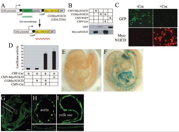
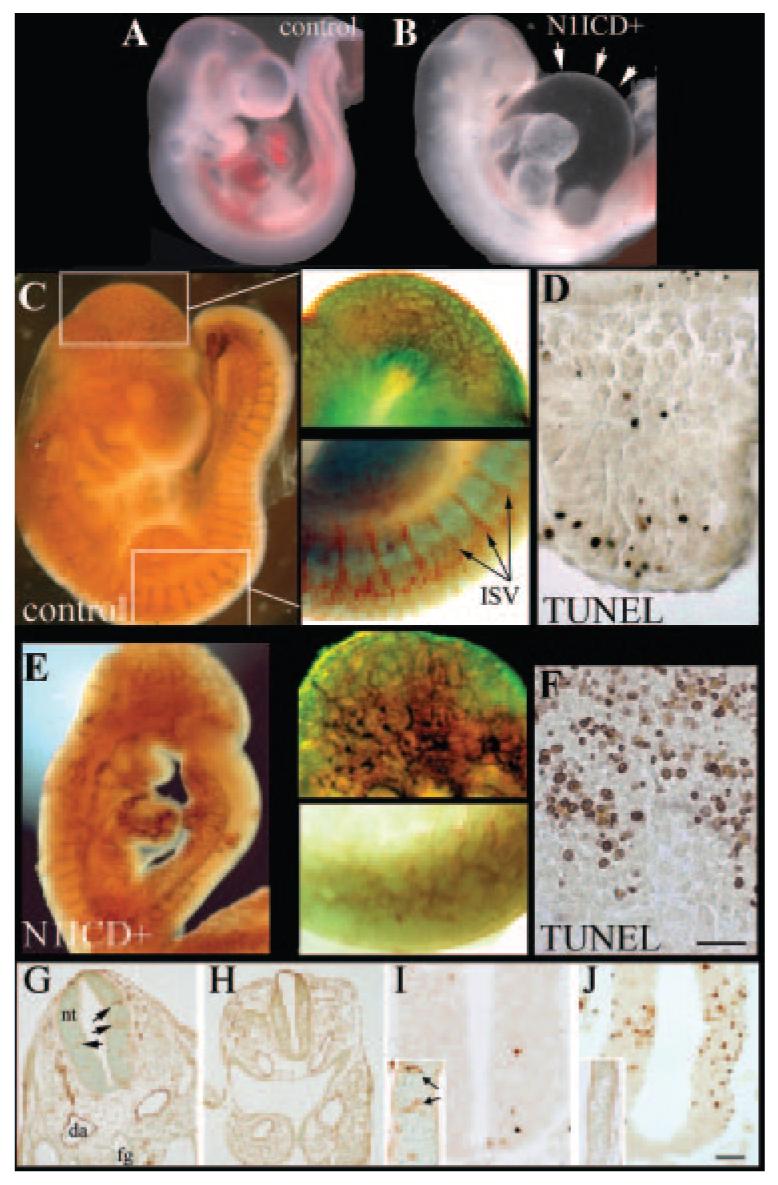
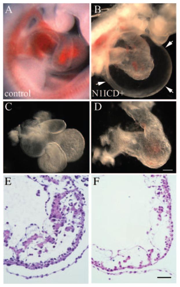
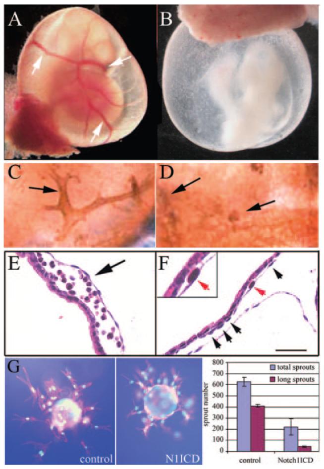
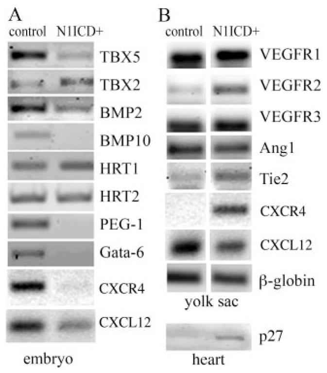
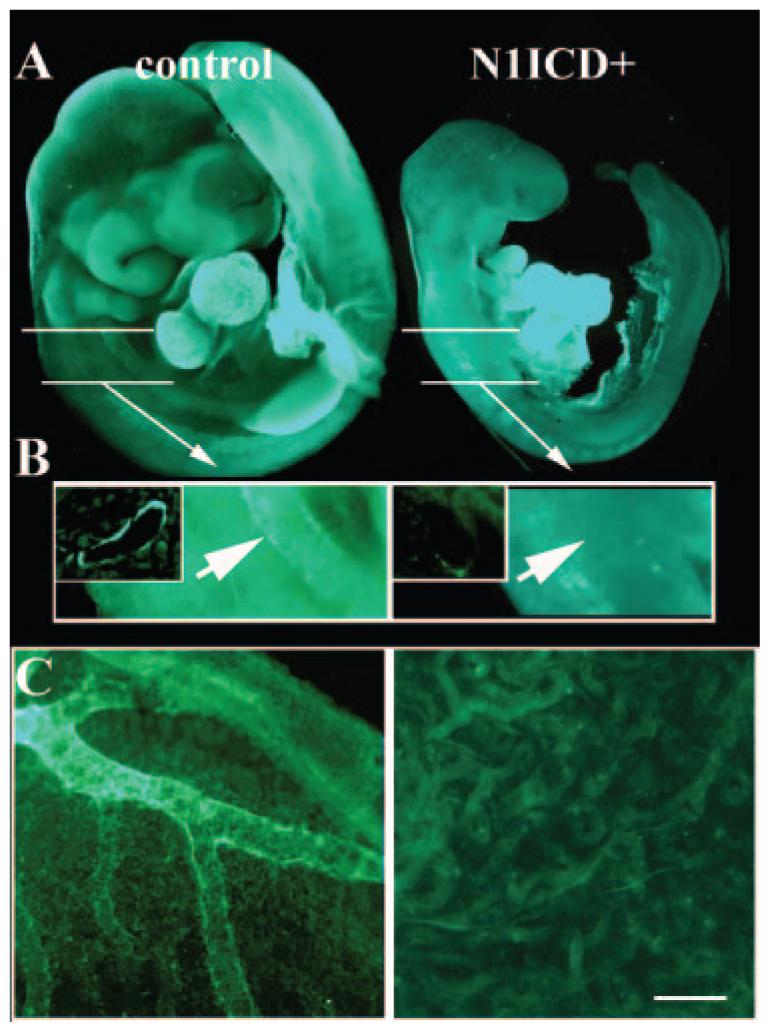
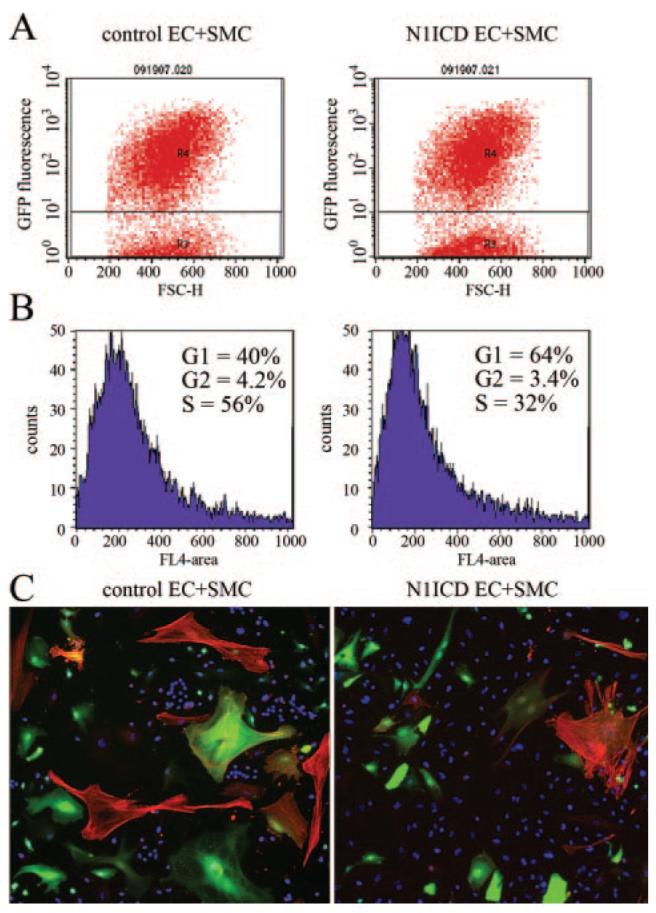
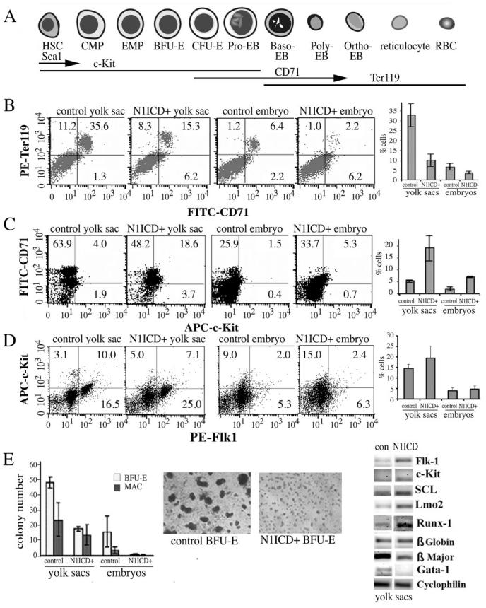
Notch signaling is critical for the development and maintenance of the cardiovasculature, with loss-of-function studies defining roles of Notch1 in the endothelial/hematopoietic lineages. No in vivo studies have addressed complementary gain-of-function strategies within these tissues to define consequences of Notch activation. We developed a transgenic model of Cre recombinase-mediated activation of a constitutively active mouse Notch1 allele (N1ICD(+)) and studied transgene activation in Tie2-expressing lineages. The in vivo phenotype was compared to effects of Notch1 activation on endothelial tubulogenesis, paracrine regulation of smooth muscle cell proliferation, and hematopoiesis. N1ICD(+) embryos showed midgestation lethality with defects in angiogenic remodeling of embryonic and yolk sac vasculature, cardiac development, smooth muscle cell investment of vessels, and hematopoietic differentiation. Angiogenic defects corresponded with impaired endothelial tubulogenesis in vitro following Notch1 activation and paracrine inhibition of smooth muscle cells when grown with Notch1-activated endothelial cells. Flow cytometric analysis of hematopoietic and endothelial precursor populations demonstrated a significant loss of CD71(+)/Ter119(+) populations with an active N1ICD(+) allele and a corresponding increase in c-Kit(+)/CD71 and Flk1(+) populations, suggesting a developmental block during the transition between c-Kit- and Ter119-expressing erythroblasts. Cardiovascular lineages are sensitive to an imbalance in Notch signaling, with aberrant activation reflecting a vascular phenotype comparable to a loss-of-function Notch1 mutation.


Figures

Figure 1
Figure 1
Development of a conditional mouse N1ICD transgene. A, The N1ICD construct is shown. B, CGMycN1ICD was transfected with or without a CMV-Cre expression plasmid in 293T cells, and expression was monitored by immunoblot (B) or immunofluorescence staining (C). CMV-MycN1ICD and CMVSGFP plasmids were used as positive controls for MycN1ICD and GFP expression, respectively. Myc fluorescence is red. D, A CBF-1—luciferase construct was cotransfected with CGMycN1ICD and CMV-Cre plasmids into 293T cells. CMVMycN1ICD plasmid was used as a positive control. Normalized luciferase activities are presented as means±SD. E and F, Tie2Cre×ROSA Cre reporter crosses were set up, and resultant negative (E) or Tie2Cre-positive (F) E9.5 embryos were prepared for detection of β-galactosidase activity. G and H, Tie2Cre×CGMycNICD crosses were obtained and embryos immunostained with anti-myc to detect the N1ICD myc tag. Positive staining was seen in embryonic vasculature (G) and endothelial cells of the dorsal aorta (H) and yolk sac (I). The area indicated in G was analyzed in the cross sections. Lower insets for H and I show background staining in control aorta and yolk sac. The scale bar in G represents 100 μm for G, 25 μm for H, and 50 μm for I.
Figure 2
Figure 2
Activation of Notch1 leads to defective embryonic cardiovasculature and lethality. Embryos from Tie2Cre× NotchICD crosses were collected at E9.5. Shown are a wild-type embryo (A and C) and a N1ICD+ littermate (B and E). N1ICD+ embryos were smaller than controls, and internal carotid artery, dorsal aorta, and aortic arch were not blood-filled. The extensive pericardial edema in N1ICD+ embryos is highlighted (B, arrows). PECAM-1 immunostaining of control (C) and N1ICD+ embryos (E) highlighted vasculature. Areas of the head and the dorsal somites shown in higher magnification (right) are boxed (C, left). Angiogenic sprouts of intersomitic vessels (ISV) were present in N1ICD+ embryos (E, lower inset), although they failed to show proper vascular patterning. This corresponded to increased TUNEL labeling of somites in N1ICD+ embryos (F) compared to controls (D). The scale bar in F represents 75 μm for the right images of C, 60 μm for the right images of E, and 50 μm for D and F. G through J, Normal (G and I) or N1ICD+ (H and J) E9.5 embryos were sectioned coronally and stained with anti-PECAM (G and H, insets in I and J) or TUNEL-labeled (I and J). N1ICD+ embryos had smaller dorsal aortae (da) and neural tubes (nt), with a lack of blood vessels within the neural tube (H) compared to control (G, arrows). Control neural tubes had minimal TUNEL-positive cells (I), corresponding to robust vascularization (inset, PECAM staining), whereas high apoptosis was observed in N1ICD+ embryos (J), consistent with a lack of vascularization (inset). The scale bar in J represents 50 μm for G and H, 12.5 μm for C and D, and 25 μm for I and J.
Figure 3
Figure 3
Heart defects in Notch1ICD+ embryos. Freshly dissected E9.5 wild-type (A) or N1ICD+ (B) embryos are shown. Isolated hearts from N1ICD+ embryos displayed abnormal ventricular looping and underdeveloped cardiac chambers (D), compared to the normally developed heart (C). The scale bar in D represents 100 μm for A and B, 70 μm for C, and 60 μm for D. E and F, Heart sections from control (E) or N1ICD+ (F) embryos were hematoxylin/eosin-stained to show ventricular trabeculation. The scale bar in F represents 25 μm for E and F.
Figure 4
Figure 4
Defects in yolk sac vasculature in Notch1ICD+ embryos. Embryos were collected from control (A, C, and E) or N1ICD+ embryos (B, D, and F) at E9.5. Whole mount views of embryos with intact yolk sacs show lack of conducting arteries (arrows, A) and orange peel—like appearance of N1ICD+ yolk sacs (B). C and D, PECAM-1-staining shows normal vascular structure (C) vs the fused primitive vascular network in N1ICD+ yolk sacs (D). E and F, hematoxylin/eosin-stained sections contrast normal blood filled vessels in control (E) with a gross enlargement between endoderm and mesoderm layers in N1ICD+ embryos (F), resulting in lacunae-like spaces. Endothelial cells were present in the N1ICD+ yolk sacs (F, arrows, inset). The scale bar in F represents 50 μm. G, Notch1ICD inhibits endothelial sprouting in vitro. Fibronectin-coated microcarrier beads were seeded with HUVEC-GFP or HUVEC-Notch1ICD, embedded in fibrin, and grown for 3 days. Normal branching was inhibited by Notch1ICD. Total sprouts and sprouts > 150 μm (long sprouts) were counted and measured and shown as means± SD.
Figure 5
Figure 5
Gene expression in transgenic N1ICD+ embryos. Semiquantitative RT-PCR analysis was performed on embryos (A) and yolk sacs (B) for expression of genes critical for hematopoiesis and angiogenesis.
Figure 6
Figure 6
Notch1 activation in Tie2 populations inhibits smooth muscle investment of vessels. Immunostaining for smooth muscle α-actin (SMA) was performed on E9.5 embryos. A, SMA expression in the developing heart was comparable in control (left) and N1ICD+ (right) embryos. White lines indicate magnified region in B. B, SMA-positive cells are identifiable surrounding the aorta in controls (arrow, left), whereas none was seen in N1ICD+ embryos (arrow, right). In the N1ICD+ embryos, some fluorescence is seen in the dorsally located somites. The insets for both images show sections through the aorta, showing SMA-positive layers in only controls. C, Yolk sacs from control (left) or N1ICD+ (right) embryos were stained for SMA. Control yolk sacs had vessels with mural cell investment, but there was no SMA staining in the rudimentary network in yolk sacs from N1ICD+ embryos (right). The scale bar in F represents 100 μm for A and 140 μm for C.
Figure 7
Figure 7
Coculture of smooth muscle cells with endothelial cells with activated Notch signaling suppresses cell proliferation. HUVECs were transduced with LacZ (control) or N1ICD adeno-virus and grown for coculture with GFP-expressing human aortic SMCs. After 3 days of coculture, cells were stained with DRAQ5, and SMCs were identified based on GFP fluorescence (A). DRAQ5-based cell cycle analysis was performed on GFP-expressing SMCs (B). C, Cultures were stained with 4′,6-diamidino-2-phenylindole (DAPI) (blue, nuclei) and SMA (red) to distinguish between SMCs (GFP and SMA) and endothelial cells (only nuclear staining).
Figure 8
Figure 8
Impaired erythroid differentiation in N1ICD+ embryos. A, Representation of erythroid differentiation and marker expression. Flow cytometry was used to analyze cell surface antigens from embryos or yolk sacs at E9.5. B, CD71+ and Ter119+ populations are shown and graphed are double CD71+/ Ter199+ populations in each group. C, CD71+ and c-Kit+ populations are shown, and graphed are the percentages of CD71+/c-Kit+ double positive cells, showing increased c-Kit+ progenitors in N1ICD+ groups. D, Analysis of Flk1+ and c-Kit+ double positive population did not show significant differences but progenitors positive for Flk1 alone were significantly increased in N1ICD+ groups (graphed). E, Methylcellulose colony-forming assays were used to quantify BFU-E and macrophage-forming colonies from yolk sacs and embryos. Shown are representative photomicrographs of BFU-E colonies 8 days after plating. E, RT-PCR was performed using primers as indicated.

References

    1. Robb L, Elefanty AG. The hemangioblast—an elusive cell captured in culture. Bioessays. 1998;20:611–614. - PubMed
    1. Risau W. Mechanisms of angiogenesis. Nature. 1997;386:671–674. - PubMed
    1. Limbourg FP, Takeshita K, Radtke F, Bronson RT, Chin MT, Liao JK. Essential role of endothelial Notch1 in angiogenesis. Circulation. 2005;111:1826–1832. - PMC - PubMed
    1. Shutter JR, Scully S, Fan W, Richards WG, Kitajewski J, Deblandre GA, Kintner CR, Stark KL. Dll4, a novel Notch ligand expressed in arterial endothelium. Genes Dev. 2000;14:1313–1318. - PMC - PubMed
    1. de Angelis M Hrabe, McIntyre J, II, Gossler A. Maintenance of somite borders in mice requires the Delta homologue DII1. Nature. 1997;386:717–721. - PubMed

Publication types

MeSH terms