. 2008 Aug;118(8):2733-46.
doi: 10.1172/JCI32381.

Ang II-stimulated migration of vascular smooth muscle cells is dependent on LR11 in mice

Affiliations

Ang II-stimulated migration of vascular smooth muscle cells is dependent on LR11 in mice

Meizi Jiang et al. J Clin Invest. 2008 Aug.

Abstract

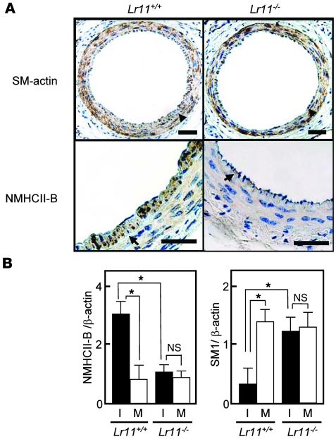
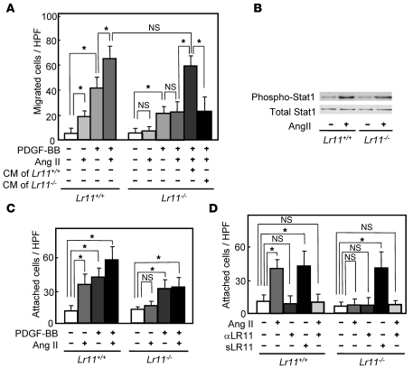
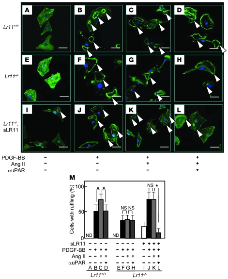
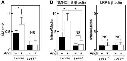
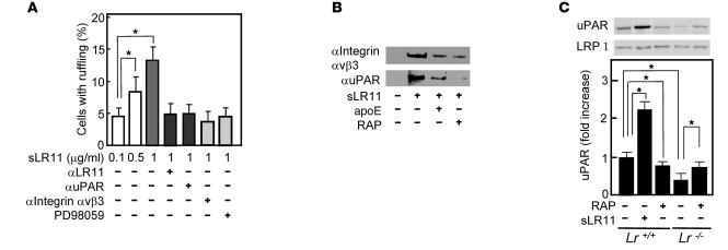
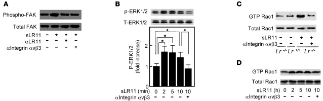
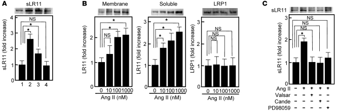
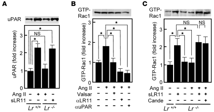
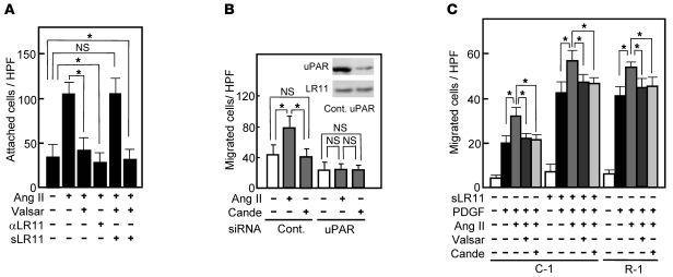
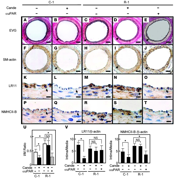
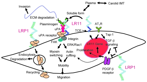
Medial-to-intimal migration of SMCs is critical to atherosclerotic plaque formation and remodeling of injured arteries. Considerable amounts of the shed soluble form of the LDL receptor relative LR11 (sLR11) produced by intimal SMCs enhance SMC migration in vitro via upregulation of urokinase-type plasminogen activator receptor (uPAR) expression. Here, we show that circulating sLR11 is a novel marker of carotid intima-media thickness (IMT) and that targeted disruption of the LR11 gene greatly reduces intimal thickening of arteries through attenuation of Ang II-induced migration of SMCs. Serum concentrations of sLR11 were positively correlated with IMT in dyslipidemic subjects, and multivariable regression analysis suggested sLR11 levels as an index of IMT, independent of classical atherosclerosis risk factors. In Lr11-/- mice, femoral artery intimal thickness after cuff placement was decreased, and Ang II-stimulated migration and attachment of SMCs from these mice were largely abolished. In isolated murine SMCs, sLR11 caused membrane ruffle formation via activation of focal adhesion kinase/ERK/Rac1 accompanied by complex formation between uPAR and integrin alphavbeta3, a process accelerated by Ang II. Overproduction of sLR11 decreased the sensitivity of Ang II-induced activation pathways to inhibition by an Ang II type 1 receptor blocker in mice. Thus, we demonstrate a requirement for sLR11 in Ang II-induced SMC migration and propose what we believe is a novel role for sLR11 as a biomarker of carotid IMT.


Figures

Figure 1
Figure 1. Intimal thickness of arteries after cuff placement in Lr11–/– mice.
(A) Targeted disruption strategy of the murine LR11 gene, consisting of 49 exons. The targeting vector (bold line) contains 3.3 kb (5′) and 4.4 kb (3′) of genomic DNA flanking the neomycin-resistance cassette (Neor). After homologous recombination, Neor replaced exon 1 (Ex1, gray box), which contained the initiation codon of the LR11 gene. The location of the probe used for Southern blot analysis is shown. RV, EcoRV; X, XbaI; H, HindIII; E, EcoRI. (B) Southern blot analysis of murine-tail DNA from heterozygous intercrosses digested with EcoRV using the probe (see Figure 1A) that detects 17-kb and 6.8-kb fragments in the WT and knockout allele, respectively. (C) Immunodetection of LR11 protein. Total protein (100 μg) extracted from brain and kidney were separated by electrophoresis, blotted on a membrane, and incubated with antibody against LR11 (~250 kDa) or LRP1(~85 kDa). The samples were loaded on the same gel but not on immediately neighboring lanes. (D) Upper panels show sections of femoral artery of Lr11+/+ or Lr11–/– mouse after cuff placement, subjected to elastica van Gieson staining. Arrowheads indicate the internal elastic layers. Scale bar: 50 μm. Lower panel shows I/M ratio of arteries presented as mean ± SD (n = 15). *P < 0.05.
Figure 2
Figure 2. Myosin isoform expression pattern in injured arteries after cuff placement in Lr11–/– mice.
(A) Sections of femoral arteries in Lr11+/+ or Lr11–/– mice after cuff placement, subjected to immunohistochemistry using antibodies against SMA or NMHCII-B. Arrowheads indicate the internal elastic layers. Scale bars: 50 μm. (B) mRNA levels of myosin isoforms NMHCII-B and SM1 in arteries after cuff placement in Lr11+/+ or Lr11–/– mice. Total RNA isolated from the thickened intima (I) or the media (M) of Lr11+/+ or Lr11–/– mice was reverse transcribed to cDNA and subjected to real-time PCR analysis using specific primers for NMHCII-B and SM1, respectively. The amounts of amplified products are expressed relative to the amounts of β-actin transcript. Data are presented as mean ± SD (n = 5). *P < 0.05.
Figure 3
Figure 3. Ang II–induced migration and attachment of cultured SMCs derived from Lr11–/– mice.
(A) Effect of Ang II on the PDGF-BB–induced (10 ng/ml) migration activities of Lr11+/+ or Lr11–/– SMCs. SMCs were incubated with 1 μM Ang II for 24 hours in the presence or absence of conditioned medium of Lr11–/– SMCs before migration analyses. Data are presented as mean ± SD (n = 6). (B) Effect of Ang II on Stat1 phosphorylation in Lr11+/+ or Lr11–/– SMCs. SMCs were incubated with 1 μM Ang II for 10 minutes before immunoblot analysis for (phospho-) Stat1 (~90 kDa). (C) Effects of Ang II on cell attachment of Lr11+/+ or Lr11–/– SMCs in the presence or absence of 10 ng/ml PDGF-BBs. SMCs were incubated with or without Ang II (1 μM) for 24 hours before attachment analyses. Data are presented as mean ± SD (n = 6). (D) Effects of sLR11 on Ang II–induced attachment of Lr11+/+ or Lr11–/– SMCs. SMCs were incubated with or without Ang II (1 μM) for 24 hours in the presence or absence of anti-LR11 antibody (pm11, 1:5 dilution) or recombinant sLR11 (1 μg/ml) for 24 hours before attachment analyses. Data are presented as mean ± SD (n = 6). *P < 0.05.
Figure 4
Figure 4. Ang II–induced membrane ruffle formation of cultured SMCs derived from Lr11–/– mice.
(AL) Ang II–induced membrane ruffling (arrowheads) in Lr11+/+ or Lr11–/– SMCs. The indicated SMCs were incubated with or without Ang II (1 μM) for 24 hours in the presence or absence of recombinant sLR11 (1 μg/ml) with or without anti-uPAR antibody for 24 hours. PDGF-BB (10 ng/ml) was then added to the culture medium for 10 minutes before immunofluorescence analyses. Cells were then stained using Alexa Fluor 488 phalloidin. Scale bars: 10 μm. (M) The number of cells with membrane ruffles were counted among 500 cells in the field. Data are presented as mean ± SD (n = 3). *P < 0.05. ND, not detected.
Figure 5
Figure 5. Intimal thickening after cuff placement in response to Ang II infusion in Lr11–/– mice.
(A) I/M ratios of femoral arteries in Lr11+/+ or Lr11–/– mice after cuff placement with saline or Ang II infusion (1 μg/kg/min for 28 days) are presented as mean ± SD (n = 15). *P < 0.05. (B) mRNA levels of NMHCII-B and LRP1 in injured arteries. Total RNA isolated from the intima or the media of Lr11+/+ or Lr11–/– mice was reverse transcribed into cDNA and subjected to real-time PCR analysis using specific primers for NMHCII-B and LRP1, respectively. The amounts of amplified products are expressed relative to the amounts of β-actin transcript, and the ratio of mRNA expression levels of intima and media are presented as mean ± SD (n = 3). *P < 0.05.
Figure 6
Figure 6. sLR11-mediated ruffle formation through complex formation with uPAR and integrin αvβ3.
(A) sLR11-induced membrane ruffle formation. Rabbit SMCs were incubated with sLR11 (1 μg/ml) for 24 hours in the presence or absence of antibody against LR11, uPAR, or integrin αvβ3 (MAB1976) or of PD98059. The number of cells with membrane ruffles were counted among 500 cells in the field. Data are presented as mean ± SD (n = 5). (B) Coimmunoprecipitation of sLR11 (~250 kDa) with integrin αvβ3 or uPAR. Membrane extracts of rabbit SMCs were incubated with or without sLR11 in the presence or absence of apoE (50 μg/ml) or RAP (10 μg/ml), immunoprecipitated with anti-integrin αvβ3 or anti-uPAR antibody, and subjected to immunoblot analysis using anti-LR11 antibody. (C) uPAR expression in Lr11–/– SMCs. Membrane extracts of Lr11+/+ or Lr11–/– SMCs were incubated with RAP and subjected to immunoblot analysis using anti-uPAR (~50 kDa) or anti-LRP1 (~85 kDa) antibody. Blot shown is representative of 3 independent experiments. Data are presented as mean ± SD (n = 3). *P < 0.05.
Figure 7
Figure 7. sLR11-mediated intracellular signals related to cytoskeleton reorganization.
(A) sLR11-induced FAK activation. Cell lysates of rabbit SMCs were incubated with or without sLR11 (1 μg/ml) in the presence or absence of antibody against LR11 or integrin αvβ3 (MAB1976), immunoprecipitated with anti-FAK antibody, and subjected to immunoblot analysis using anti-FAK (~130 kDa) or anti–phospho-FAK (~130 kDa) antibody. (B) sLR11–induced phosphorylation of ERK1/2. Cell lysates (10 μg protein) of rabbit SMCs were incubated with sLR11 (1 μg/ml) for the indicated times in the presence or absence of anti–integrin αvβ3 antibody (MAB1976) and subjected to immunoblot analysis using antibody against (phospho) p42/44 MAP kinase. Upper and lower signals represent ERK1 (~44 kDa) and ERK2 (~42 kDa), respectively (13). Blot shown is representative of 3 independent experiments. Data of p-ERK1/2 are presented as mean ± SD (n = 3). *P < 0.05. (C) Rac1 activation in Lr11–/– SMCs. Cell lysates (60 μg protein) of Lr11+/+ or Lr11–/– SMCs were incubated with sLR11 (1 μg/ml) in the presence or absence of antibody against integrin αvβ3 (RMV-7), immunoprecipitated with PAK-1 PBD Protein GST beads, and subjected to immunoblot analysis with anti-Rac1 (~21 kDa) or anti–GTP-Rac1 (~21 kDa) antibody. (D) sLR11-induced Rac1 activation. Cell lysates (60 μg protein) of rabbit SMCs were incubated with sLR11 (1 μg/ml) for the indicated times in the presence or absence of anti–integrin αvβ3 antibody (MAB1976) and subjected to immunoblot analysis with anti-Rac1 (Rac1 ~21 kDa and GST-Rac1 ~21 kDa) antibody with (top) or without (bottom) prior immunoprecipitation with PAK-1 PBD Protein GST beads.
Figure 8
Figure 8. Ang II–induced sLR11 production in SMCs.
(A) Effects of chemotactic cytokines on the production of sLR11 in rabbit SMCs. Conditioned media collected for 12 hours in the absence (lane 1) or presence of Ang II (1 μM, lane 2), PDGF (10 ng/ml, lane 3), or VEGF (50 ng/ml, lane 4) were concentrated and subjected to immunoblot analysis using anti-LR11 antibody (~250 kDa). Blot shown is representative of 3 independent experiments. Data are presented as mean ± SD (n = 3). *P < 0.05. (B) Ang II–dependent increase of soluble or membrane-bound forms of LR11 in SMCs. Membrane extracts (20 μg protein) prepared from rabbit SMCs or conditioned medium collected for 12 hours after the addition of Ang II at the indicated concentrations were subjected to immunoblot analysis with anti-LR11 (~250 kDa) or anti-LRP1 (~85 kDa) antibody. Blot shown is representative of 3 independent experiments. Data are presented as mean ± SD (n = 3). (C) Effect of ARBs or an ERK inhibitor on the Ang II–dependent increase in sLR11 in rabbit SMCs. Conditioned medium collected for 12 hours in the presence or absence of Ang II with or without valsartan (valsar, 10 nM), candesartan (10 nM), or PD98059 (10 μM) were subjected to immunoblot analysis with anti-LR11 (~250 kDa) antibody. Blot shown is representative of 3 independent experiments. Data are presented as mean ± SD (n = 3).
Figure 9
Figure 9. Ang II–induced LR11/uPAR pathway in SMCs.
(A) uPAR expression in Lr11–/– SMCs. Membrane extracts of Lr11+/+ SMCs or Lr11–/– SMCs were incubated with or without Ang II (1 μM) in the presence or absence of sLR11 (1 μg/μl) and subjected to immunoblot analysis using anti-uPAR (~50 kDa) antibody. Blot shown is representative of 3 independent experiments. Data are presented as mean ± SD (n = 3). *P < 0.05. (B) Effect of blocking the LR11/uPAR pathway on the Ang II–dependent increase of Rac1 activation in rabbit SMCs. Cell lysates (60 μg protein) were incubated in the presence or absence of Ang II (1 μM) with or without valsartan (10 nM), anti-LR11 antibody, or anti-uPAR antibody, immunoprecipitated with PAK-1 PBD Protein GST beads, and subjected to immunoblot analysis using anti-Rac1 (~21 kDa) antibody. Blot shown is representative of 3 independent experiments. Data are presented as mean ± SD (n = 3). (C) Effect of ARB on the Ang II–dependent increase of Rac1 activation in Lr11–/– SMCs. Cell lysate (60 μg protein) of Lr11+/+ or Lr11–/– SMCs was incubated in the presence or absence of Ang II (1 μM) with or without candesartan (10 nM) or sLR11 (1 μg/μl), immunoprecipitated with PAK-1 PBD Protein GST beads, and subjected to immunoblot analysis using anti-Rac1 (~21 kDa) antibody. Blot shown is representative of 3 independent experiments. Data are presented as mean ± SD (n = 3).
Figure 10
Figure 10. Ang II–induced migration via the LR11/uPAR pathway in SMCs.
(A) Effect of blocking LR11 activation on the Ang II–induced increase of attachment of rabbit SMCs. The SMCs attached to plastic plates were counted after incubation in the presence or absence of Ang II (1 μM) with or without valsartan (10 nM), anti-LR11 antibody, or sLR11 (1 μg/μl). Data are presented as mean ± SD (n = 3). *P < 0.05. (B) Effect of uPAR silencing on the Ang II–induced increase in rabbit SMCs migration. The migrated SMCs treated with exogeneous siRNA specific for uPAR or control (Cont.) RNA were counted after incubation in the presence or absence of Ang II (1 μM) with or without candesartan (10 nM). Inset: Membrane extracts (10 μg protein) prepared from these cells were subjected to immunoblot analysis with anti-uPAR (~50 kDa) or anti-LR11 antibody (~250 kDa). Data are presented as mean ± SD (n = 3). (C) Effect of Ang II on the PDGF-induced migration of LR11-overexpressing SMCs. The number of migrated A7r5 cells transfected with LR11 cDNA (R-1), or control cells (C-1) in the presence or absence of PDGF-BB (PDGF, 10 ng/ml) were determined after incubation with or without sLR11 (1 μg/μl), Ang II (1 μM), or valsartan (10 nM). Data are presented as mean ± SD (n = 10).
Figure 11
Figure 11. Effect of the ARB candesartan on intimal thickness after arterial injury in sLR11-overproducing mice.
BL6 nude mice were implanted subcutaneously with A7r5 cells transfected with LR11 cDNA (R-1) or control cells (C-1). Sections of femoral arteries of cell-implanted mice after cuff placement with or without administration of candesartan or anti-uPAR neutralizing antibody were subjected to histological analysis using elastica van Gieson (EVG) staining (AE). Serial sections were immunohistochemically analyzed using antibody against SMA (FJ), LR11 (KO), or NMHCII-B (PT). Arrowheads indicate the internal elastic layers. Scale bars: 50 μm (AJ); 10 μm (KT). (U) I/M ratio of arteries is presented as mean ± SD (n = 5). *P < 0.05. (V) mRNA levels of LR11 and NMHCII-B in injured arteries. Total RNA isolated from thickened intima or media of mice using LCM was reverse transcribed and subjected to real-time PCR analysis using specific primers for NMHCII-B and LR11, respectively. The amounts of amplified products are expressed relative to the amounts of β-actin transcript, and the ratio of mRNA expression levels of intima and media are presented as mean ± SD (n = 3).
Figure 12
Figure 12. Proposed molecular mechanism for LR11 requirement in the response of SMCs to Ang II.
Ang II and PDGF-BB are the key cytokines promoting migration of SMCs in plaque formation. LRP1 inhibits the PDGF-BB–mediated signals for migration and proliferation and/or the modulation of upstream Tsp-1/TGF-β–mediated signals (38) through interaction with the PDGF-β receptor. Ang II induces the Tsp-1/TGF-β signals (37) and LR11 gene transcription through activating AT1R. LR11 localized on the cell surface becomes the soluble form (as sLR11) by cleavage through TNF-α–converting enzyme (TCE) (22). Circulating sLR11 levels are positively correlated with carotid IMT. sLR11 binds to and interacts with uPAR, the expression of which is mainly regulated by LRP1, on the cell surface and/or on neighboring cells. This complex formation inhibits the internalization of uPAR via LRP1, resulting in enhanced uPAR cell-surface expression. The uPA/uPAR system increases cell mobility through both increased ECM degradation and intracellular integrin/FAK/ERK/Rac-1 signaling, which in turn promotes actin ruffling and myosin isoform switching. The SMCs expressing LR11 display increased migratory capacity in response to PDGF-BB and/or Ang II. Thus, LR11 in combination with its counteracting partner LRP1 regulates the migration of intimal SMCs in injured arteries and atherosclerotic plaques via modulation of the uPA/uPAR system. The proposed LR11-mediated migration of intimal SMCs may be modulated by other Ang II–induced molecules and cytokines, particularly endothelial cell–derived PAI-1 (48). ARBs inhibit (a) the migration of intimal SMCs through downregulation of LR11 and (b) their proliferation by blockade of signals mediated by Tsp-1/TGF-β and PDGF-BB.

References

    1. Ross R. Atherosclerosis — an inflammatory disease. N. Engl. J. Med. . 1999;340:115–126. doi: 10.1056/NEJM199901143400207. - DOI - PubMed
    1. Newby A.C. Dual role of matrix metalloproteinases (matrixins) in intimal thickening and atherosclerotic plaque rupture. Physiol. Rev. 2005;85:1–31. doi: 10.1152/physrev.00048.2003. - DOI - PubMed
    1. Blasi F., Carmeliet P. uPAR: a versatile signalling orchestrator. Nat. Rev. Mol. Cell. Biol. 2002;3:932–943. doi: 10.1038/nrm977. - DOI - PubMed
    1. Reuning U., Magdolen V., Hapke S., Schmitt M. Molecular and functional interdependence of the urokinase-type plasminogen activator system with integrins. Biol. Chem. 2003;384:1119–1131. doi: 10.1515/BC.2003.125. - DOI - PubMed
    1. Herz J., Hui D.Y. Lipoprotein receptors in the vascular wall. Curr. Opin. Lipidol. 2004;15:175–181. doi: 10.1097/00041433-200404000-00010. - DOI - PubMed

Publication types