Review
. 2008 Jul;15(7):665-74.
doi: 10.1038/nsmb.1450.

Synaptic vesicle fusion

Affiliations
Review

Synaptic vesicle fusion

Josep Rizo et al. Nat Struct Mol Biol. 2008 Jul.

Abstract

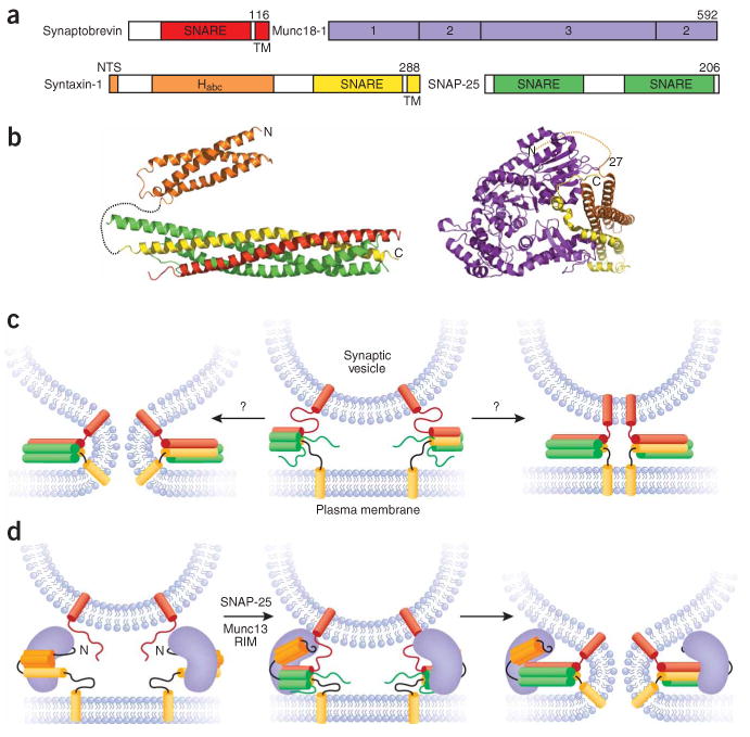
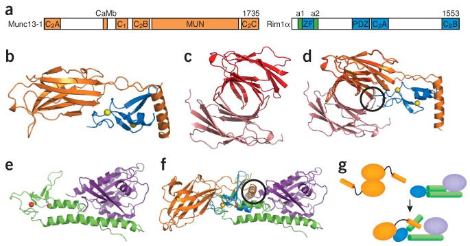
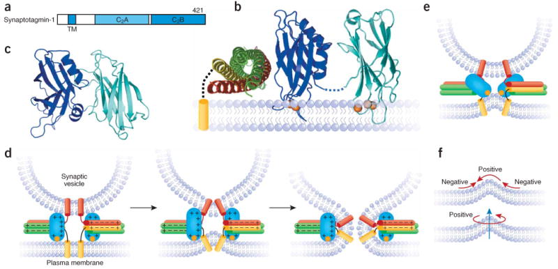
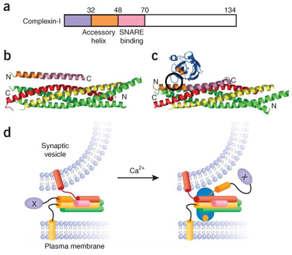
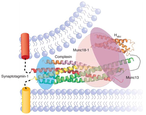
The core of the neurotransmitter release machinery is formed by SNARE complexes, which bring the vesicle and plasma membranes together and are key for fusion, and by Munc18-1, which controls SNARE-complex formation and may also have a direct role in fusion. In addition, SNARE complex assembly is likely orchestrated by Munc13s and RIMs, active-zone proteins that function in vesicle priming and diverse forms of presynaptic plasticity. Synaptotagmin-1 mediates triggering of release by Ca2+, probably through interactions with SNAREs and both membranes, as well as through a tight interplay with complexins. Elucidation of the release mechanism will require a full understanding of the network of interactions among all these proteins and the membranes.


Figures

Figure 1
Figure 1
SNAREs and Munc18-1. (a) Domain diagrams of the neuronal SNAREs and Munc18-1. The number of residues of each protein is indicated above each diagram on the right. The same color coding for these proteins is used in all other figures. TM, transmembrane. (b) Ribbon diagrams of the structures of the SNARE complex and the syntaxin-1 Habc domain (shown connected by a dashed curve that represents the syntaxin-1 sequence linking them) and of the binary complex between Munc18-1 and the closed conformation of syntaxin-1. The orange dashed curve indicates that the syntaxin-1 NTS also participates in Munc18-1 binding,, even though it was not observed in the structure of the complex. The N and C termini of syntaxin-1 are indicated in both diagrams. (c) Partially assembled SNARE complexes (center) and how they could induce membrane fusion as they fully assemble (left) or could remained fully assembled between the two membranes (right) if the linkers between the SNARE motifs and transmembrane regions are flexible. (d) The closed conformation of syntaxin-1 bound to Munc18-1 (left) and Munc18-1 bound to partially assembled SNARE complexes (center) that could induce fusion as the SNARE complexes fully assemble (right). Munc13s and RIMs are likely to mediate the transition between the two complexes.
Figure 2
Figure 2
Munc13s and RIMs. (a) Domain diagram of Munc13-1 and RIM1α. The number of residues of each protein is indicated above each diagram on the right. The helices adjacent to the RIM1α ZF domain, which are involved in Rab3 binding, are labeled a1 and a2. The same color-coding is used in b,dg. (b) Structure of the complex between the RIM2α ZF domain and the N-terminal region of Munc13-1, including the C2A domain. Yellow spheres, zinc ions. (c,d) Structure of the Munc13-1 C2A domain homodimer (c) and superposition with the Munc13-1/RIM2α heterodimer (d). The two protomers of the Munc13-1 C2A domain homodimer are shown in red and salmon to distinguish them from the Munc13-1 C2A domain in the heterodimer. (e) Structure of Rab3A (purple) bound to the rabphilin N-terminal region containing its ZF domain and the two adjacent helices involved in Rab3 binding (green). Red spheres, zinc ions. (f) Superposition of the Munc13-1–RIM2α and Rab3A–rabphilin structures shown in b and e using the two ZF domains for the superposition. Black circles, regions of overlap between the RIM2α ZF domain and one Munc13-1 C2A domain protomer of the homodimer in d, and between Rab3A and the Munc13-1 α-helix at the C terminus of the C2A domain in f. (g) Model summarizing the structural rearrangements and changes in protein-protein interfaces proposed to occur during the switch from the Munc13-1 homodimer to the Munc13-1–RIM-Rab3 tripartite complex.
Figure 3
Figure 3
Syntaptotagmin-1 and its coupling to SNAREs and membranes. (a) Domain structure of synaptotagmin-1, with the number of residues indicated in the top right corner. TM, transmembrane. (b) Model of the SSCAP complex built from the structures of the SNARE complex and the Ca2+-bound synaptotagmin-1 C2 domains, and a mutagenesis analysis of SSCAP complex formation. Orange spheres, Ca2+ ions; dashed black curve, the linker between the syntaxin-1 SNARE motif and transmembrane region. Note that it is uncertain whether the synaptotagmin-1 Ca2+-binding loops bind to the plasma membrane or the synaptic vesicle membrane. (c) Crystal structure of the tandem synaptotagmin-1 C2 domains in the absence of Ca2+; the structure involves an antiparallel interaction between the two domains that needs to be disrupted to allow Ca2+ binding to the C2A domain. (d) Model of how the synaptotagmin-1 C2B domain could cooperate with the SNAREs in triggering membrane fusion upon Ca2+ influx by binding to both membranes and the C terminus of the SNARE complex. Each orange circle represents the two Ca2+ ions bound to the C2B domain. The + and − signs illustrate the electrostatic charge distribution of the C2B domain and the SNARE complex. The model assumes that synaptotagmin-1 interacts with the SNARE complex through the polybasic region on the side of the C2B domain, as in b. This interaction is weak in solution but is likely to be strengthened by colocalization of synaptotagmin-1 and the SNARE complex on one membrane, which at the same time may increase binding specificity by disfavoring irrelevant interactions existing in solution between these highly charged molecules. The C2A domain is not shown in this model for simplicity, but could play a related role. (e) Model of how the Ca2+ binding loops of the synaptotagmin-1 C2 domains (only the C2B domain is shown for simplicity) could help to cause membrane fusion by inducing positive curvature on the plasma membrane. (f) Two diagrams showing the types of curvature involved in membrane bending and illustrating that such bending requires both positive and negative curvature.
Figure 4
Figure 4
Complexins and their coupling to SNAREs and synaptotagmin-1. (a) Domain diagram of complexin-I. Residue numbers are indicated above the diagram. The same color-coding is used in the remaining panels. (b) Structure of a complexin-I fragment bound to the SNARE complex. The N and C termini of the complexin-I fragment and the SNARE complex are indicated. (c) Superposition of the crystal structure shown in b with the model of the SCCAP complex shown in Figure 3b (omitting the membrane and the C2A domain). Black circle, the region of overlap between the C2B domain and the accessory helix of complexin-I. (d) Models of how binding of the accessory helix to the C terminus of the SNARE complex could hinder full SNARE complex assembly and thus inhibit fusion (left), and how this inhibition could be released by partial competition between the synaptotagmin-1 C2B domain and complexin for binding to the C terminus of the SNARE complex (right). The N terminus of complexin is shown as a purple ellipse with an X in the center to indicate an as-yet-unidentified interaction of this region that is critical for complexin function. The orientation of this N-terminal region in both diagrams is arbitrary.
Figure 5
Figure 5
Diagram illustrating the notion that Munc18-1, Munc13, complexin and synaptotagmin-1 bind to the SNARE complex. The ribbon diagram shows the structure of the complexin-I–SNARE complex, which illustrates the only one among these interactions that has been revealed at atomic resolution. The binding site of synaptotagmin-1 on the four-helix bundle is inferred from mutagenesis, whereas those of Munc18-1 and Munc13 are unknown. The figure intends to illustrate that all these SNARE complex interactions may be compatible, mutually exclusive, or partially competitive, and that resolving this issue as well as how these interactions occur during the steps that lead to exocytosis will be critical to understand the mechanism of release.

References

    1. Sudhof TC. The synaptic vesicle cycle. Annu Rev Neurosci. 2004;27:509–547. - PubMed
    1. Rosenmund C, Rettig J, Brose N. Molecular mechanisms of active zone function. Curr Opin Neurobiol. 2003;13:509–519. - PubMed
    1. Brunger AT. Structure and function of SNARE and SNARE-interacting proteins. Q Rev Biophys. 2005;38:1–47. - PubMed
    1. Rizo J, Chen X, Arac D. Unraveling the mechanisms of synaptotagmin and SNARE function in neurotransmitter release. Trends Cell Biol. 2006;16:339–350. - PubMed
    1. Jahn R, Scheller RH. SNAREs — engines for membrane fusion. Nat Rev Mol Cell Biol. 2006;7:631–643. - PubMed

Publication types

MeSH terms