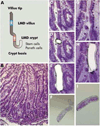
Defining disease with laser precision: laser capture microdissection in gastroenterology
- PMID: 18619446
- PMCID: PMC3736118
- DOI: 10.1053/j.gastro.2008.06.054
Defining disease with laser precision: laser capture microdissection in gastroenterology
Abstract
Laser capture microdissection (LCM) is an efficient and precise method for obtaining pure cell populations or specific cells of interest from a given tissue sample. LCM has been applied to animal and human gastroenterology research in analyzing the protein, DNA, and RNA from all organs of the gastrointestinal system. There are numerous potential applications for this technology in gastroenterology research, including malignancies of the esophagus, stomach, colon, biliary tract, and liver. This technology can also be used to study gastrointestinal infections, inflammatory bowel disease, pancreatitis, motility, malabsorption, and radiation enteropathy. LCM has multiple advantages when compared with conventional methods of microdissection, and this technology can be exploited to identify precursors to disease, diagnostic biomarkers, and therapeutic interventions.
Conflict of interest statement
Figures


References
-
- Emmert-Buck MR, Bonner RF, Smith PD, Chuaqui RF, Zhuang Z, Goldstein SR, Weiss RA, Liotta LA. Laser capture microdissection. Science. 1996;274:998–1001. - PubMed
-
- Stappenbeck TS, Hooper LV, Manchester JK, Wong MH, Gordon JI. Laser capture microdissection of mouse intestine: characterizing mRNA and protein expression, and profiling intermediary metabolism in specified cell populations. Methods Enzymol. 2002;356:167–196. - PubMed
-
- Streitz JM, Jr, Madden MT, Marimanikkuppam SS, Krick TP, Salo WL, Aufderheide AC. Analysis of protein expression patterns in Barrett's esophagus using MALDI mass spectrometry, in search of malignancy biomarkers. Dis Esophagus. 2005;18:170–176. - PubMed
-
- Melle C, Bogumil R, Ernst G, Schimmel B, Bleul A, von Eggeling F. Detection and identification of heat shock protein 10 as a biomarker in colorectal cancer by protein profiling. Proteomics. 2006;6:2600–2608. - PubMed
Publication types
MeSH terms
Substances
Grants and funding
LinkOut - more resources
Full Text Sources

