. 2008 Jul 22;18(14):1072-7.
doi: 10.1016/j.cub.2008.06.050. Epub 2008 Jul 10.

Osmotic regulation of Rab-mediated organelle docking

Affiliations

Osmotic regulation of Rab-mediated organelle docking

Christopher L Brett et al. Curr Biol. .

Abstract

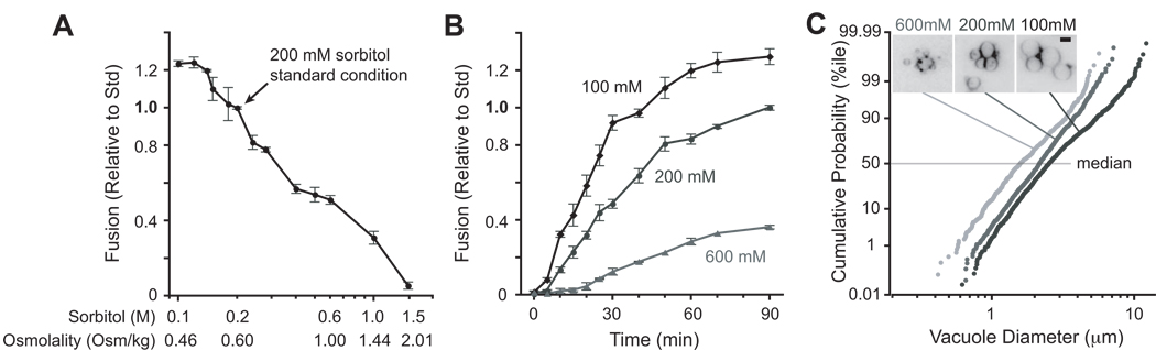
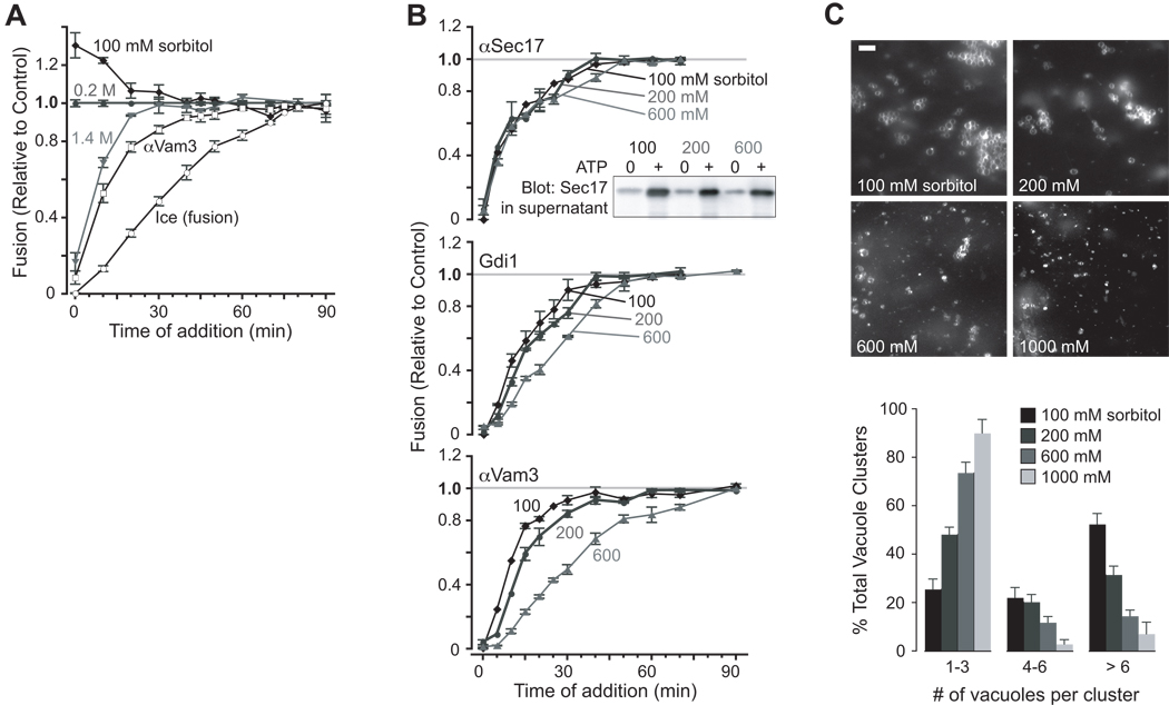
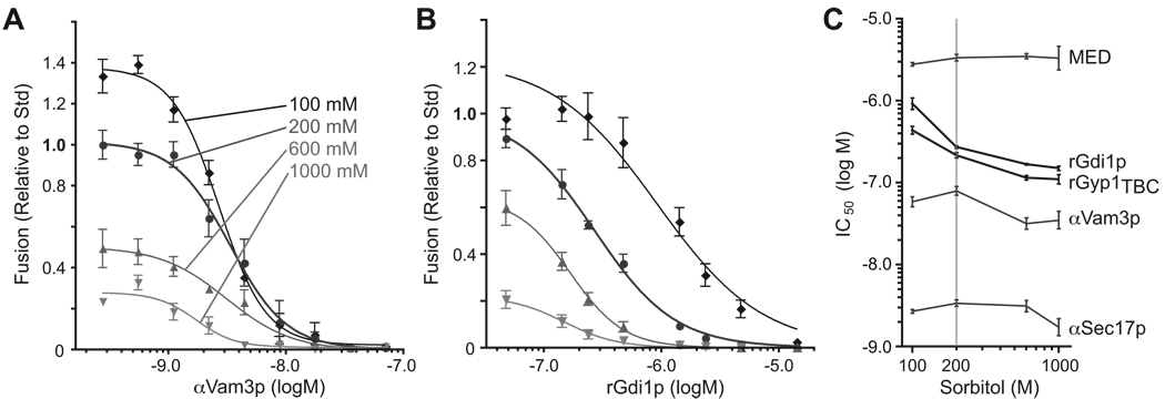
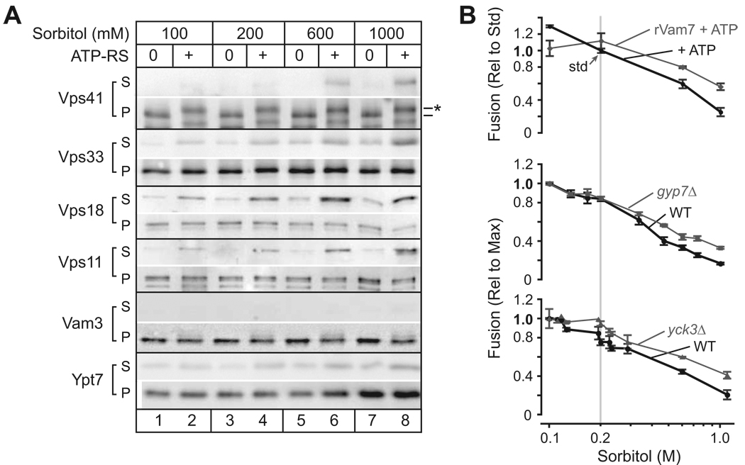
Osmotic gradients across organelle and plasma membranes modulate the rates of membrane fission and fusion; sufficiently large gradients can cause membrane rupture [1-6]. Hypotonic gradients applied to living yeast cells trigger prompt (within seconds) swelling and fusion of Saccharomyces cerevisiae vacuoles, whereas hypertonic gradients cause vacuoles to fragment on a slower time scale [7-11]. Here, we analyze the influence of osmotic strength on homotypic fusion of isolated yeast vacuoles. Consistent with previously reported in vivo results, we find that decreases in osmolyte concentration increase the rate and extent of vacuole fusion in vitro, whereas increases in osmolyte concentration prevent fusion. Unexpectedly, our results reveal that osmolytes regulate fusion by inhibiting early Rab-dependent docking or predocking events, not late events. Our experiments reveal an organelle-autonomous pathway that may control organelle surface-to-volume ratio, size, and copy number: Decreasing the osmolyte concentration in the cytoplasmic compartment accelerates Rab-mediated docking and fusion. By altering the relationship between the organelle surface and its enclosed volume, fusion in turn reduces the risk of membrane rupture.


Figures

Figure 1
Figure 1. Osmotic control of homotypic vacuole fusion
(A) In vitro homotypic vacuole fusion at the indicated concentrations of sorbitol was measured after a 90 min. incubation. On the horizontal axis, measured buffer osmolality is shown for reaction buffers containing the indicated concentrations of sorbitol. n≥4 for each point. (B) Kinetics of fusion in hypotonic (100 mM), standard (200 mM), and hypertonic (600 mM sorbitol) conditions. n ≥ 4 for each point; bars span 95% confidence intervals. (C) Vacuole diameter increases with fusion. Fusion increases both membrane area and volume, and hence the size of the vacuoles in the population. To crosscheck the results obtained in content mixing assays, vacuole diameter was measured at the end of 70 min. fusion reactions containing the indicated concentrations of sorbitol. The morphometric data are plotted in rank order in cumulative probability histograms., wiht the median vacuole diameter (50th %ile) for each treatment indicated. 500–1000 vacuoles from at least two experiments were measured for each condition. (Inset) Examples of morphology of FM4-64-stained vacuoles after 70 min. incubation with ATP. Scale bar = 2.5 µm.
Figure 2
Figure 2. Osmotic control of early docking subreactions
(A) Kinetic mapping of osmotic effect. At the times indicated, aliquots of standard vacuole fusion reactions (200 mM sorbitol) were shifted to a final sorbitol concentration of 100, 200 or 1400 mM, as indicated, and incubated for a total of 90 min. Data are normalized to the values obtained with reactions diluted in isotonic (200 mM sorbitol) buffer. Kinetics are also shown for inhibitors of docking (αVam3p antibody, 35 nM) and fusion (ice) at standard 200 mM sorbitol. n ≥ 4 independent experiments. (B) Osmotic control of docking kinetics. At the indicated times, subreaction inhibitors were added to reactions containing 100, 200 or 600 mM sorbitol throughout the incubation. Fusion inhibitors include (from early to late stage inhibition): 322 nM αSec17 antibody; 14 µM rGdi1; 35 nM αVam3 antibody. The data are normalized to fusion values obtained when fusion buffer was added in place of inhibitor at the indicated times (grey lines). (Inset) Vacuoles were incubated with or without ATP for 10 min. in the presence of 100, 200 or 600 mM sorbitol, then sedimented. The amount of Sec17 released from the membrane pellet was determined by western blot analysis using 1/6th of the supernatant from each fusion reaction. n ≥ 4 independent experiments. (C) Osmotic inhibition of tethering. Images of FM4-64 stained vacuoles after 30 min. of incubation with or without ATP in the presence of 100, 200, 600 or 1000 mM sorbitol. Scale bar = 6 µm. Vacuole clusters were scored under each condition, and data is presented as a percentage of total. A cluster refers to a single group of vacuoles. At least 200 clusters were counted for each condition.
Figure 3
Figure 3. Osmolytes enhance sensitivity of fusion to Rab inhibitors
Homotypic vacuole fusion was measured after vacuoles were incubated with ATP for 90 min. in the presence of 100, 200, 600 or 1000 mM sorbitol and increasing concentrations of (A) anti-Vam3 antibody or (B) rGdi1p. Sigmoidal dose-response curves were fit to the datasets. (C) IC50 values were extracted from the fits shown in A and B, and from similar curves for other inhibitors (see Fig. S3A–C). Bars span 95% confidence intervals; n ≥ 4 for all experiments shown. Dose-response for additional inhibitors and an alternative presentation of the IC50 values is presented in Fig. S3D.
Figure 4
Figure 4. Interplay between osmotic strength and docking factors
(A) Osmolyte-triggered HOPS dissociation from the vacuole. Vacuoles were incubated with or without ATP for 70 min. in the presence of 100, 200, 600 or 1000 mM sorbitol, then sedimented. Membrane association of 4 HOPS components (Vps41, Vps33, Vps18 and Vps11), a vacuole Qa-SNARE (Vam3), and the vacuole Rab GTPase (Ypt7) was assessed by western blot using 1/3 of the total isolated supernatent (S) and 1/10 (0.6 µg vacuole protein) of the pellet (P). Phospho-Vps41 (*) was observed exclusively in the presence of ATP, as reported [11]. (B) Rab activation or partial Rab bypass attenuates inhibition by osmolyte. Top: Vacuoles were incubated for 90 min. with ATP at increasing sorbitol concentrations in the the presence and absence of recombinant Qc-SNARE rVam7, which reduces the Ypt7 requirement in fusion [21, 22], Middle, bottom: vacuoles were purified from yeast strains containing or lacking Gyp7 (middle), which when absent stabilizes membrane association of Ypt7 and components of HOPS (inset B; Fig. 4S), or the vacuolar casein kinase I Yck3 (bottom), which, when absent, stabilizes HOPS on the membrane [11]. Fusion data were normalized to either the mean rate of fusion under standard conditions (ATP only, 200 mM sorbitol; top) or conditions where maximum fusion values were observed (100 mM sorbitol; middle, bottom). n ≥ 4 for each point.

References

    1. Hogue MJ. The effect of hypotonic and hypertonic solutions on fibroblasts of the embryonic chick heart in vitro. J Exp Med. 1919;30:617–648. - PMC - PubMed
    1. Tominaga T, Allen RD, Naitoh Y. Cyclic changes in the tension of the contractile vacuole complex membrane control its exocytotic cycle. J Exp Biol. 1998;201:2647–2658. - PubMed
    1. Zimmerberg J, Sardet C, Epel D. Exocytosis of sea urchin egg cortical vesicles in vitro is retarded by hyperosmotic sucrose: kinetics of fusion monitored by quantitative light-scattering microscopy. J Cell Biol. 1985;101:2398–2410. - PMC - PubMed
    1. Dai J, Ting-Beall HP, Sheetz MP. The secretion-coupled endocytosis correlates with membrane tension changes in RBL 2H3 cells. J Gen Physiol. 1997;110:1–10. - PMC - PubMed
    1. Fatt P, Katz B. Spontaneous subthreshold activity at motor nerve endings. J Physiol. 1952;117:109–128. - PMC - PubMed

Publication types

MeSH terms

Substances

LinkOut - more resources